ABSTRACT
- Lipopolysaccharide (LPS), which consists of a lipid A anchor, a core oligosaccharide, and a highly variable O-antigen, plays an important role in maintaining membrane integrity and preventing harmful extracellular molecules from penetrating the membrane of Gram-negative bacteria. Although the roles of lipid A and O-antigen have been extensively studied, the role of the core oligosaccharide in envelope stress responses and antibiotic resistance remains relatively poorly understood. In this study, we analyzed the roles of ten enzymes involved in core biosynthesis and modification in envelope stress responses and antibiotic resistance. Our systematic analyses demonstrated that biosynthesis and modification of heptoses present in the inner core of LPS are important for resistance to bile salts and antibiotics. Notably, we found that phosphorylation of the first heptose in the inner core had a role distinct from that of phosphorylation of the second heptose. Defects in heptose biosynthesis resulted in increased membrane permeability. Finally, we found that SpeA/SpeB-dependent putrescine biosynthesis may be partially associated with the sensitivity of the heptose biosynthesis-defective mutant to clindamycin and erythromycin. Taken together, our results demonstrate that the biosynthesis and modification of heptoses in the inner core of LPS are strongly associated with the envelope stress response and antibiotic resistance.
-
Keywords: Lipopolysaccharide, core, heptose, envelope stress response, antibiotic resistance, bile salts, lipopolysaccharide modification
Introduction
The outer membrane of Gram-negative bacteria has an asymmetric bilayer architecture, with an inner leaflet composed of phospholipids and an outer leaflet composed of lipopolysaccharide (LPS) (Sun et al., 2022). LPS is specific to Gram-negative bacteria and is composed of hydrophobic saturated acyl chains and various hydrophilic sugars (Mitchell and Silhavy, 2019). These structural features prevent the passage of both hydrophilic and hydrophobic molecules across the outer membrane (Mitchell and Silhavy, 2019; Sun et al., 2022). LPS consists of lipid A containing a glucosamine disaccharide, a core oligosaccharide, and O-antigen polysaccharide chains (VanOtterloo et al., 2024). Lipid A is a glucosamine disaccharide with predominantly six acyl chains, with occasional instances of more than six, and two phosphate groups (Hummels, 2025). These structural features of lipid A influence the innate immune response and resistance to colistin and other polymyxins (Gogry et al., 2021; Raetz and Whitfield, 2002).
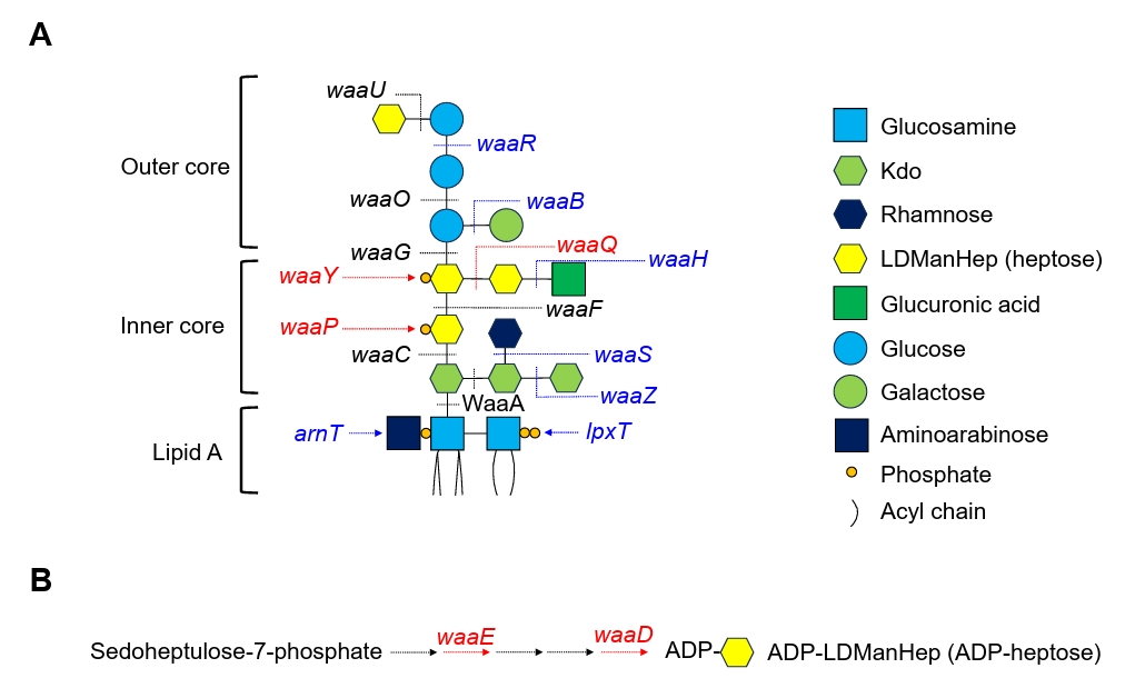
The core region of LPS consists of an inner core oligosaccharide composed of 3-deoxy-D-manno-octulosonic acid (Kdo) and L-glycero-D-manno-heptose (hereafter, heptose), as well as an outer core oligosaccharide composed of glucose, galactose, and heptose (Di Lorenzo et al., 2022). The composition of core oligosaccharides varies considerably across bacterial species (VanOtterloo et al., 2024). In Escherichia coli, the inner core oligosaccharide comprises three Kdo residues and three heptoses; in some cases, glucuronic acid or rhamnose may also be added (Fig. 1). The outer core oligosaccharide comprises three glucoses, one galactose, and one heptose. The first and second heptoses in the inner core oligosaccharide are phosphorylated by the heptose-specific kinases WaaP and WaaY, respectively (Yethon and Whitfield, 2001; Yethon et al., 1998), whereas the third heptose is not phosphorylated (Fig. 1). Phosphorylation of sugar moieties in lipid A and the inner core substantially increases the hydrophilicity of LPS. Although the effects of lipid A on the innate immune response and antibiotic resistance have been extensively studied (Gogry et al., 2021; Raetz and Whitfield, 2002), the role of the inner or outer core oligosaccharides in envelope stress responses and antibiotic resistance remains relatively poorly understood.
In this study, we analyzed the roles of the inner and outer core oligosaccharides in the envelope stress response and antibiotic resistance. Our systematic analyses showed that heptoses in the inner core oligosaccharide are important for resistance to bile salts and various antibiotics, including clindamycin and novobiocin. Loss of either WaaD or WaaE, both of which are involved in heptose biosynthesis, resulted in increased membrane permeability. Through random mutagenesis, we showed that the inactivation of SpeA/SpeB-dependent putrescine biosynthesis partially alleviated the sensitivity of the waaE mutant to clindamycin and erythromycin. Additionally, we found that phosphorylation of the first heptose had a role distinct from that of phosphorylation of the second heptose. Taken together, our results demonstrate that the biosynthesis and modification of heptoses in the inner core are strongly associated with the envelope stress response and antibiotic resistance.
Materials and Methods
Bacterial strains, plasmids, and culture conditions
All E. coli strains and plasmids used in this study are listed in Table S1. All deletion mutant strains were constructed using λ red recombinase expressed from the pKD46 plasmid, as previously described (Datsenko and Wanner, 2000; Son et al., 2024). Deletion cassettes containing a kanamycin resistance gene and an FRT sequence were amplified from the pKD13 plasmid by polymerase chain reaction (PCR) using primers containing 50-bp homologous sequences (Table S2). After purification, the amplified DNA fragments were introduced into E. coli MG1655 cells by electroporation. Deletion mutants were identified among the colonies that grew on kanamycin-containing Luria–Bertani (LB) agar plates and were confirmed by PCR using a separate primer set (Table S2). The kanamycin resistance gene was removed by recombination between the FRT sequences using FLP recombinase expressed from the pCP20 plasmid. To construct pACYC184-WaaD and pACYC184-WaaE, the entire open reading frames of waaD and waaE genes, together with their respective promoter regions, were amplified by PCR using genomic DNAs from MG1655 cells as a template. After purification, the amplified DNA fragments were inserted into pACYC184 digested with BamHI and EagI using In-Fusion cloning (Clontech, USA), as described previously (Ryu et al., 2025). The constructions of pACYC184-WaaD and pACYC184-WaaE plasmids were confirmed by PCR and sequencing of the PCR products. All bacterial strains were cultured in LB medium at 37°C unless otherwise stated. To prepare the alkaline LB medium, Tris was added to a final concentration of 50 mM, and the pH was adjusted using 10 N NaOH. To prepare the acidic LB medium, sodium citrate was added to a final concentration of 50 mM, and the pH was adjusted using 10 N HCl.
Determination of minimal inhibitory concentrations (MICs)
The MICs of the antibiotics against E. coli MG1655 and mutant strains were determined according to the guidelines provided by the Clinical and Laboratory Standards Institute (CLSI, 2024). Bacterial cells cultured overnight in LB medium were inoculated into Mueller–Hinton (MH) broth and cultured to a turbidity equivalent to a 0.5 McFarland standard (approximately 1.5 × 10⁸ cells/ml). The cultured cells were diluted to a final concentration of 107 cells/ml, and 10 μl of each diluted sample was spotted onto MH agar plates containing 2-fold serial dilutions of the antibiotics at final concentrations ranging from 0.0625 to 1,024 μg/ml. The MH plates were incubated at 37°C for 20 h. The MIC of each antibiotic was defined as the lowest antibiotic concentration that inhibited a full lawn growth of cells. The MIC of each antibiotic was determined by two independent experiments.
Extraction and staining of LPS
LPS was extracted using ethylenediaminetetraacetic acid (EDTA) and Triton X-100, as described previously (Lee et al., 2015). Cells cultured to stationary phase in 10 ml of LB medium were harvested in a 1.5-ml tube. The harvested cells were boiled for 15 min in a solution containing 10 mM Tris-Cl (pH 8.0), 2% Triton X-100, and 50 mM MgCl2. After cooling for 10 min at room temperature, the samples were centrifuged at 16,000 × g for 15 min at 25°C. The insoluble LPS was solubilized by incubation in a solution containing 2% Triton X-100 and 50 mM EDTA for 4 h at 37°C. The samples were then centrifuged at 16,000 × g for 15 min at 37°C. The resulting supernatant was transferred to a fresh 1.5-ml tube. The LPS in the supernatant was precipitated by the addition of MgCl2 to a final concentration of 150 mM. After incubation for 3 h at 37°C, the mixtures were centrifuged at 20,000 × g for 90 min at 37°C. The transparent LPS precipitate in the pellet was solubilized in 2× sodium dodecyl sulfate (SDS) sample buffer. After boiling for 5 min, the solubilized LPS was separated by SDS-PAGE and visualized by silver staining.
Examination of membrane permeability
Bacterial membrane permeability was assessed using chlorophenyl red-β-D-galactopyranoside (CPRG) and SYTOX green. CPRG, a lactose derivative, penetrates cells only when membrane permeability is increased. Intracellular β-galactosidase then hydrolyzes CPRG to produce chlorophenyl red, which results in red-colored colonies (Paradis-Bleau et al., 2014). SYTOX green is a membrane-impermeable fluorescent DNA dye that stains cells with increased membrane permeability (Roth et al., 1997). Wild-type and mutant cells were spotted onto LB agar plates containing 20 μg/ml CPRG. After incubation for 20 h at 37°C, photographs of the plates were taken using a digital camera EOS 100D (Canon Inc., Japan). For SYTOX green staining, overnight cultures were inoculated into fresh LB medium. Cells in the exponential phase (OD600 = 1.0) were stained with 0.5 μM SYTOX green. The stained cells were spotted onto a 1% agarose pad prepared in phosphate-buffered saline and examined under a Nikon Eclipse Ni microscope (Nikon, Japan). Cells exhibiting fluorescence intensity more than fivefold higher than the background were defined as SYTOX green-positive cells.
Random mutagenesis using mini-Tn5 transposon
Random mutagenesis was performed using the pir-dependent transposon-delivery plasmid pRL27, which contains a mini-Tn5 transposon with a kanamycin resistance gene and a transposase gene, as described previously (Choi et al., 2025; Larsen et al., 2002; Lee et al., 2021). Because pRL27 can replicate only in cells carrying the pir gene, the kanamycin resistance gene of mini-Tn5 can be maintained in E. coli MG1655 cells only when the mini-Tn5 transposon has inserted into the chromosome. Plasmid pRL27 was extracted from DH5αλpir cells carrying the pir gene and incorporated into MG1655 ΔwaaE cells by electroporation. MG1655 ΔwaaE cells carrying transposon insertions were spread onto LB agar plates containing 10 μg/ml clindamycin and 50 μg/ml kanamycin. The transposon insertion sites in the isolated suppressors were amplified by PCR using a mini-Tn5 transposon inner primer and an arbitrary primer containing a random sequence and a GGCGGT sequence (Table S2). After purification, the PCR products were sequenced using a second mini-Tn5-specific inner primer (Table S2).
Results
Inner core heptoses in LPS are important for bile salt resistance
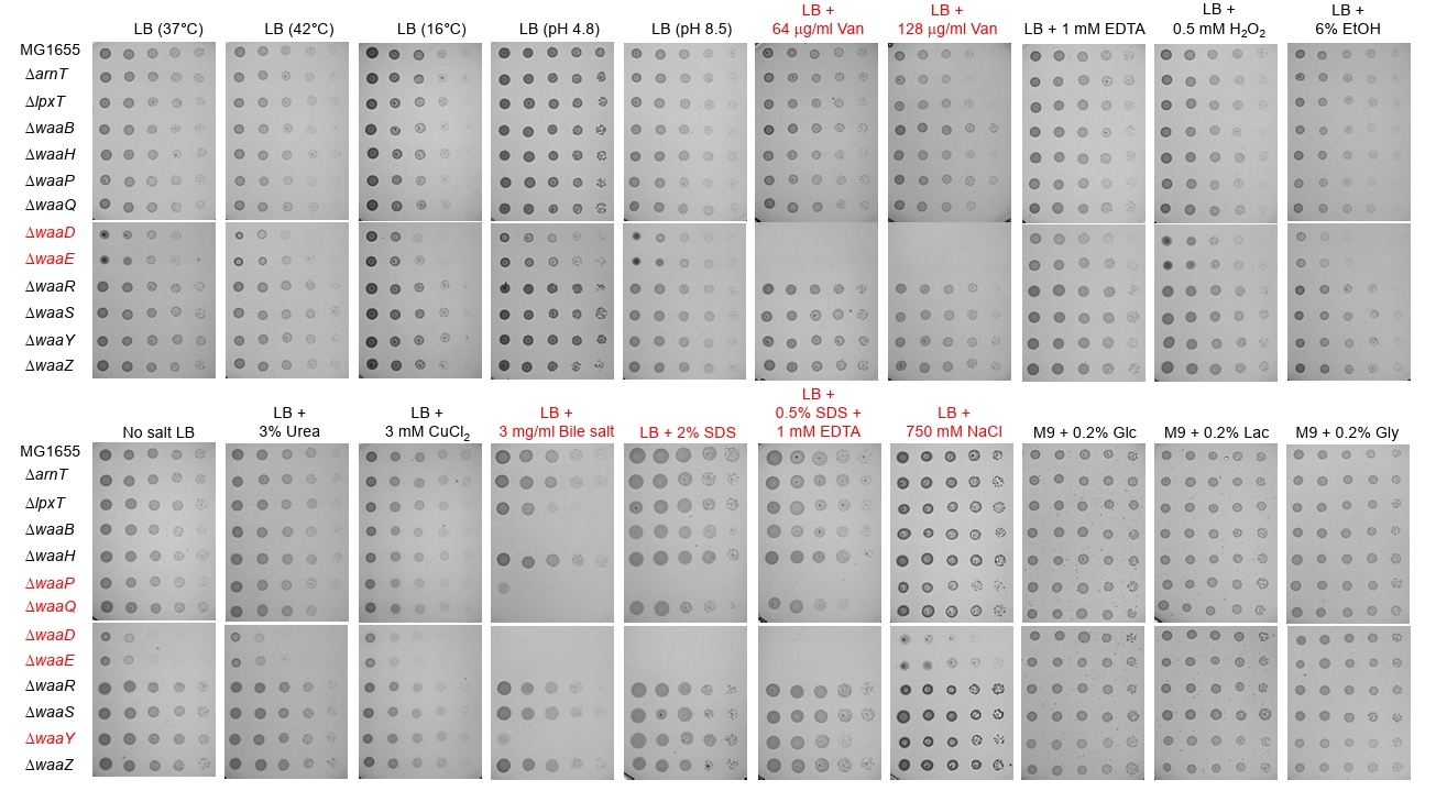
To determine the roles of LPS core sugars in the envelope stress response and antibiotic resistance, we generated mutant strains with defects in 10 non-essential genes involved in LPS core-sugar biosynthesis or modification (Fig. 1, blue- or red-colored genes). As controls, we generated mutants lacking either of two enzymes (LpxT and ArnT) involved in lipid A modification (Figs. 1 and S1). We assessed the growth of these mutant strains under various envelope stress conditions. Notably, defects in the inner core heptose biosynthesis and phosphorylation markedly increased sensitivity to various envelope stresses, including bile salts (Figs. 1 and 2, red colors). WaaD/HldD and WaaE/HldE are involved in the biosynthesis of ADP-heptose, a precursor of inner core heptose (Kneidinger et al., 2002) (Fig. S2). WaaQ is a heptosyltransferase responsible for the addition of a third heptose to the inner core of LPS (Yethon et al., 1998). WaaP and WaaY are heptose-specific kinases that phosphorylate the first and second heptose residues, respectively, in the inner core of LPS (Yethon et al., 1998; Yethon and Whitfield, 2001). All mutants lacking these enzymes exhibited marked sensitivity to bile salts (Fig. 2). The waaP, waaD, and waaE mutants exhibited marked sensitivity to SDS and SDS/EDTA stresses, whereas the waaY mutant did not (Fig. 2). In addition, the waaD and waaE mutants exhibited marked sensitivity to salt and vancomycin, whereas the other mutants did not (Fig. 2), suggesting that these enzymes have distinct roles in the envelope stress response. Collectively, these results indicate that inner core heptoses in LPS are important for resistance to various envelope stresses, including bile salts.
Disruption of inner core heptose biosynthesis or modification increases susceptibility to various antibiotics
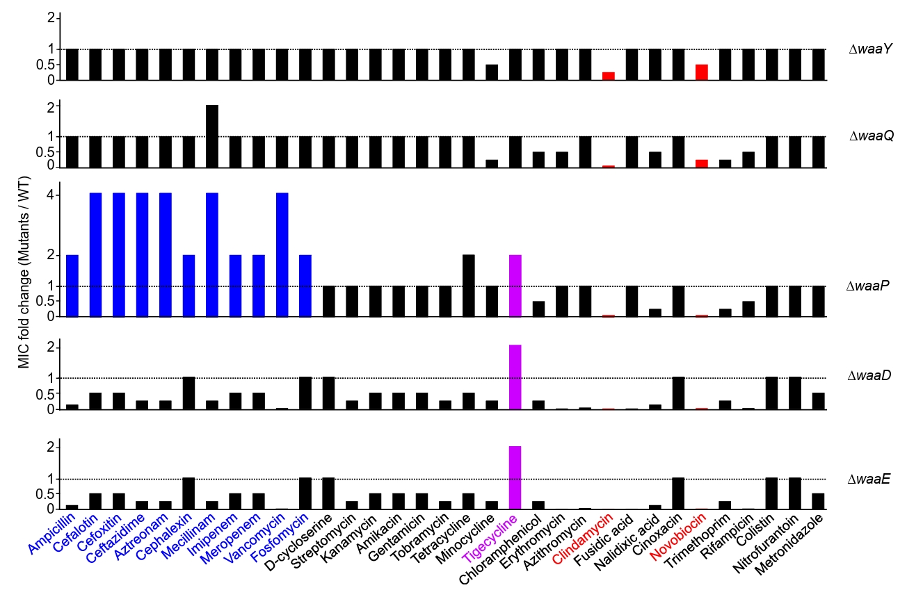
We next examined whether antibiotic susceptibility differs between the mutant and wild-type strains. The MICs of various antibiotics were measured in the wild-type and 12 mutant strains (Figs. 3 and S3). All mutant strains with defects in inner core heptose biosynthesis or modification exhibited increased susceptibility to clindamycin and novobiocin (Fig. 3, red bars). In particular, the MICs of clindamycin and novobiocin were 256-fold or 64-fold lower, respectively, in both the waaD and waaE mutants than in the wild-type strain. In both waaD and waaE mutants, the MICs of erythromycin (128-fold), fusidic acid (128-fold), vancomycin (64-fold), and rifampicin (64-fold), also strongly decreased (Fig. 3). Notably, although the MICs of most antibiotics decreased or remained unchanged in the waaD and waaE mutants, the MIC of tigecycline was modestly increased in both mutants than in the wild-type strain (Fig. 3, purple bars). The MIC of tigecycline was also modestly increased in the waaP mutant. The MICs of all tested antibiotics that target peptidoglycan biosynthesis, including β-lactams, vancomycin, and fosfomycin, were also modestly increased in the waaP mutant (Fig. 3, blue bars). Notably, these increases were not observed in the mutant lacking WaaY (Fig. 3), another heptose-specific kinase (Fig. 1), which suggests that phosphorylation of the first inner core heptose might be associated with resistances to peptidoglycan-associated antibiotics. Taken together, these results demonstrate that disruption of inner core heptose biosynthesis or modification increases susceptibility to various antibiotics, such as clindamycin and novobiocin. However, certain defects in inner core heptose biosynthesis or modification reduced susceptibility to several antibiotics, such as tigecycline and peptidoglycan-associated antibiotics.
Loss of WaaD or WaaE, which are involved in heptose biosynthesis, increases membrane permeability
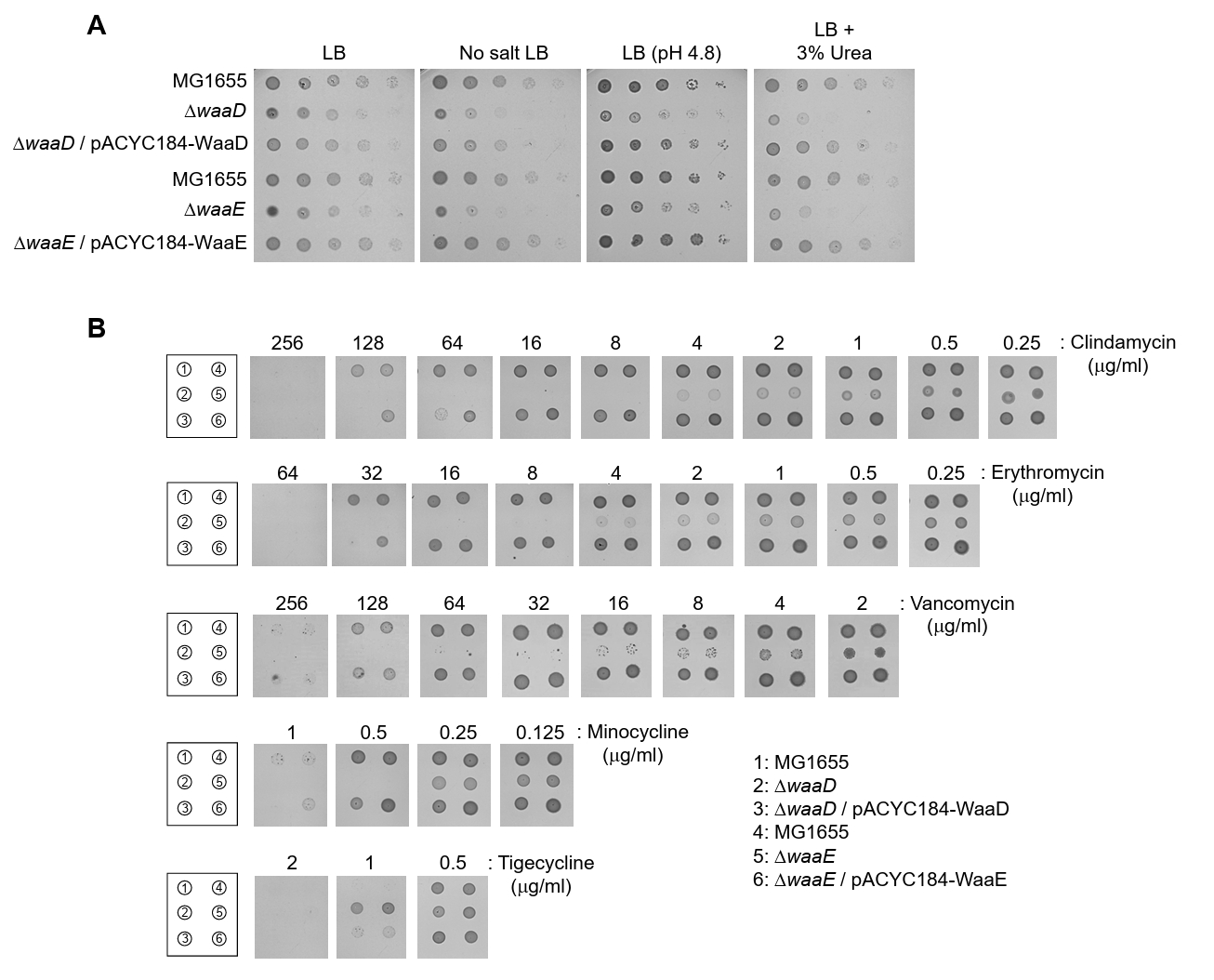
Because the most dramatic phenotypic changes were observed in the waaD and waaE mutants (Figs. 2 and 3), we focused on these mutants in further experiments. To determine whether ectopic expression of the respective genes could complement the phenotypes of the waaD and waaE mutants, we constructed two pACYC184-based plasmids that express WaaD or WaaE. The growth defects of the waaD and waaE mutants under envelope stress were almost completely complemented by pACYC184 plasmid-based expression of WaaD and WaaE, respectively (Figs. 4A and S4). Similarly, ectopic expressions of WaaD and WaaE almost completely restored antibiotic susceptibility in the waaD and waaE mutants, respectively (Fig. 4B). These results indicate that the observed phenotypes result from deletion of the respective genes.
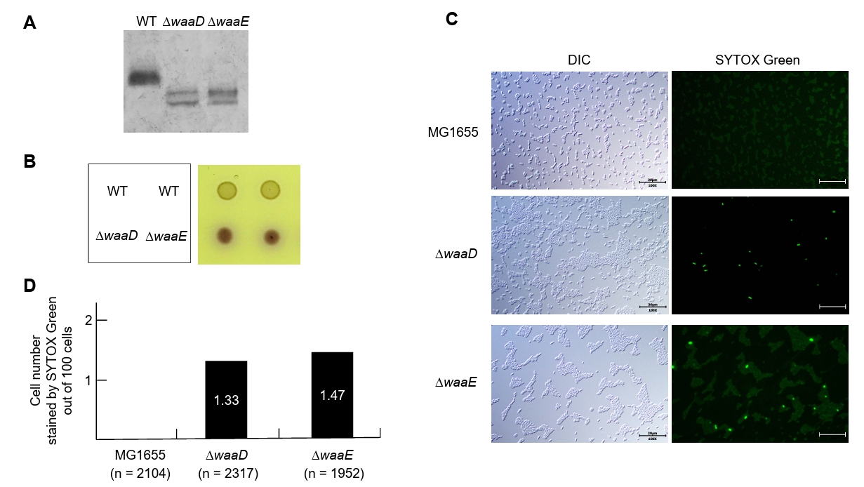
Deletion of either waaD or waaE gene is expected to result in the production of truncated LPS (Fig. 1). To confirm this, we extracted LPSs from wild-type and mutant strains. As expected, the waaD and waaE mutants produced truncated LPS compared with the wild-type strain (Fig. 5A). We next examined whether the production of truncated LPS is associated with increased membrane permeability. CPRG, a lactose derivative, enters cells with increased membrane permeability and is subsequently hydrolyzed by intracellular β-galactosidase to produce chlorophenyl red, which results in red-colored colonies (Paradis-Bleau et al., 2014). The waaD and waaE mutants formed red-colored colonies in the presence of CPRG, whereas the wild-type strain did not (Fig. 5B). Membrane permeability was also estimated using SYTOX green, a cell-impermeable fluorescent DNA dye (Langsrud and Sundheim, 1996; Roth et al., 1997). SYTOX green staining reflects compromised cell envelope integrity rather than specifically measuring outer membrane permeability. No SYTOX green fluorescence was observed in the wild-type strain, whereas strong fluorescence was detected in a subset of cells from the waaD and waaE mutants (Fig. 5C and 5D). Collectively, these results demonstrate that the production of truncated LPS in the waaD and waaE mutants is associated with increased membrane permeability. Thus, the increased membrane permeability may have contributed to the various phenotypes of the waaD and waaE mutants.
Inactivation of SpeA/SpeB-dependent putrescine biosynthesis partially alleviates the sensitivity of the waaE mutant to clindamycin and erythromycin
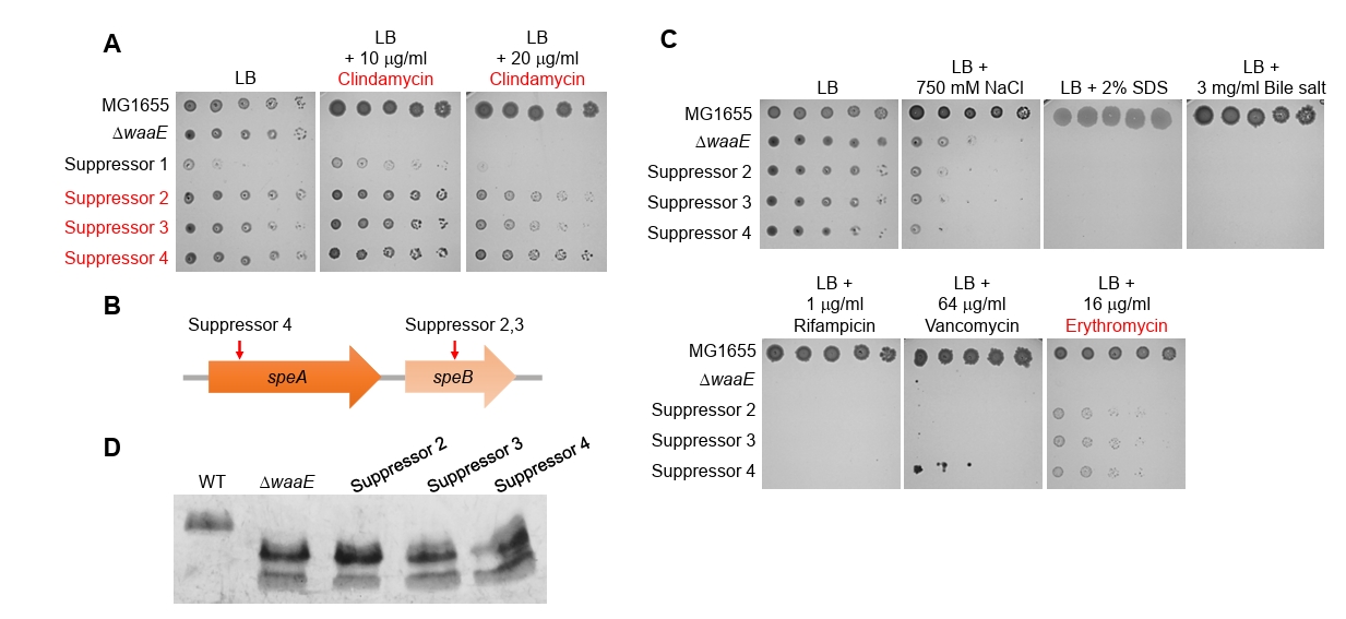
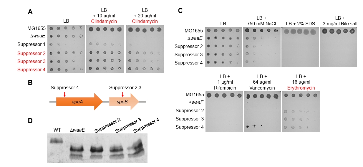
To examine the clindamycin sensitivity of the waaD and waaE mutants in more detail, we used mini-Tn5 transposon mutagenesis to isolate four mutants that partially suppressed clindamycin sensitivity of the waaE mutant. Because suppressor 1 weakly alleviated the clindamycin sensitivity of the waaE mutant and exhibited severely impaired growth in LB medium (Fig. 6A), the transposon insertion sites were mapped only in the other three suppressors. Notably, all mini-Tn5 transposon insertions were located within speA and speB (Fig. 6B), which encode enzymes involved in the biosynthesis of putrescine, a positively charged polyamine that interacts with nucleic acids, stimulates enzymatic activity, and stabilizes the membrane (Gevrekci, 2017; Huang et al., 1990; Tabor and Tabor, 1985). We next investigated whether these suppressors could restore the other phenotypes of the waaE mutant. Most of the tested phenotypes were not restored by these suppressors; however, the erythromycin sensitivity of the waaE mutant was slightly reduced (Fig. 6C). Because most phenotypes of the waaE mutant were not restored by these suppressors, we hypothesized that they did not restore the normal LPS structure. As expected, all suppressors exhibited no apparent restoration of the wild-type LPS electrophoretic pattern (Fig. 6D), indicating that their partially restored growth in the presence of clindamycin or erythromycin did not result from restoration of full-length LPS. Collectively, these results suggest that SpeA/SpeB-dependent putrescine biosynthesis may partially contribute to the sensitivity of the waaE mutant to clindamycin and erythromycin.
Discussion
LPS constitutes the first barrier encountered by external substances that attempt to enter the cells of Gram-negative bacteria. Therefore, LPS modification can substantially affect membrane permeability of external substances. However, the effects of LPS core modification on membrane permeability remain poorly understood. In this study, we investigated how the modification of LPS core components affects the envelope stress response and antibiotic resistance. Our systematic analyses revealed that the biosynthesis and modification of inner core heptoses in LPS are important for resistance to bile salts and antibiotics. Defects in the biosynthesis of inner core heptoses resulted in increased membrane permeability (Fig. 5). These results suggest that proper biosynthesis and modification of inner core heptoses are necessary to restrict the penetration of external molecules, such as bile salts and antibiotics.
In E. coli, three heptoses are attached to the Kdo residues, and the first and second heptoses are phosphorylated by the heptose-specific kinases WaaP and WaaY, respectively (Fig. 1). Our study demonstrates that both WaaP and WaaY are associated with resistance to bile salts and antibiotics. Notably, the phenotypic effects associated with the loss of these two heptose-specific kinases differed significantly. The waaP mutant did not grow under SDS or EDTA/SDS stress, whereas the waaY mutant exhibited no growth defect under these conditions (Fig. 2). The MICs of both clindamycin and novobiocin were 16-fold lower in the waaP mutant than in the wild-type strain, whereas their MICs were 4-fold or 2-fold lower, respectively, in the waaY mutant (Fig. 2). Interestingly, the MICs of peptidoglycan-associated antibiotics, such as all tested β-lactams, vancomycin, and fosfomycin, modestly increased in the waaP mutant; however, these increases were not observed in the waaY mutant (Fig. 3). The molecular mechanisms underlying these distinct phenotypes of the waaP and waaY mutants remain unclear. Future studies should clarify these underlying mechanisms.
The production of truncated LPS by the waaD and waaE mutants (Fig. 5A), was associated with increased membrane permeability (Fig. 5B, 5C, and 5D). Consistent with their increased membrane permeability, the MICs of most antibiotics were lower in these mutants (Fig. 3). However, the MIC of tigecycline was modestly increased in both the waaD and waaE mutants than in the wild-type strain (Fig. 3). Notably, the MIC of tigecycline was also modestly increased in the waaP mutant (Fig. 3). To our knowledge, the relationship between tigecycline susceptibility and inner core heptoses in LPS has not been reported. Therefore, further studies should investigate the mechanism underlying the increased MIC of tigecycline in these mutants.
We demonstrated that the inactivation of SpeA/SpeB-dependent putrescine biosynthesis partially reduced hypersensitivity of the waaE mutant to clindamycin and erythromycin, but did not alleviate its other phenotypes (Fig. 6). Clindamycin and erythromycin are lincosamide and macrolide antibiotics, respectively, that inhibit bacterial protein synthesis by blocking ribosomal translocation (Lin et al., 2018). Despite their different chemical structures, these antibiotics bind to adjacent sites on the large ribosomal subunit (50S in bacteria) and share similar resistance mechanisms (Chen et al., 2023; Lin et al., 2018; Vester and Douthwaite, 2001). Therefore, if putrescine affects ribosomal activity, particularly translocation, disruption of SpeA/SpeB-dependent putrescine biosynthesis may influence resistance to clindamycin and erythromycin. Because intracellular putrescine levels were not measured in this study, further studies are needed to determine whether changes in intracellular putrescine levels are associated with altered ribosomal activity.
Several chemical inhibitors of WaaE have been identified (De Leon et al., 2006; Desroy et al., 2009; Durka et al., 2011). Because the waaE mutant exhibited increased susceptibility to clindamycin, erythromycin, fusidic acid, rifampicin, novobiocin, and vancomycin (Fig. 3), WaaE inhibitors may potentiate the antibacterial activity of these antibiotics. This issue should be addressed in future studies.
Acknowledgments
This work was supported by a research grant from Basic Science Research Program through the National Research Foundation of Korea funded by the Ministry of Education (NRF-RS-2026-25483497) and a study on the “Gyeonggi Regional Innovation System & Education Project (Gyeonggi RISE Project)”, supported by the Ministry of Education and Gyeonggi Province (2025-RISE-09-A15).
Conflict of Interest
The authors have no financial conflicts of interest to declare.
Supplementary Information
The online version contains supplementary material available at https://doi.org/10.71150/jm.2609012
Fig. S3.
The effects of deletions of genes involved in the biosynthesis and modification of core on antibiotic susceptibility. The MIC values of the indicated antibiotics against the wild-type and indicated mutant strains were determined in MH medium. The relative MICs of the mutant strains were calculated using the MIC of the wild-type strain as the reference.
jm-2609012-Supplementary-Fig-S3.pdf
Fig. S4.
Growth defects of the waaD and waaE mutants harboring the empty pACYC184 plasmid under envelope stresses. The indicated cells were serially diluted 10-fold from 108 to 104 cells/ml and spotted onto an LB agar plate with or without the indicated compounds. No salt LB indicates LB agar plates without NaCl.
jm-2609012-Supplementary-Fig-S4.pdf
Fig. 1.Schematic representation of the roles of genes involved in the biosynthesis of LPS and heptose. (A) Schematic representation of the roles of genes involved in LPS biosynthesis. The LPS structure of the relevant E. coli K-12 was presented. The MG1655 strain lacks O-antigen under standard conditions. The inner core region consists of three Kdo residues and three heptoses. The outer core region contains three glucoses, one galactose, and one heptose. Dashed lines indicate the genes encoding the enzymes that catalyze the transfer of each sugar or phosphate moiety. Red colored genes (genes associated with the transfer or modification of heptose) and blue colored genes were deleted in this study. (B) Simplified schematic representation of the roles of genes involved in heptose biosynthesis. The waaD and waaE genes (red color) were deleted in this study.
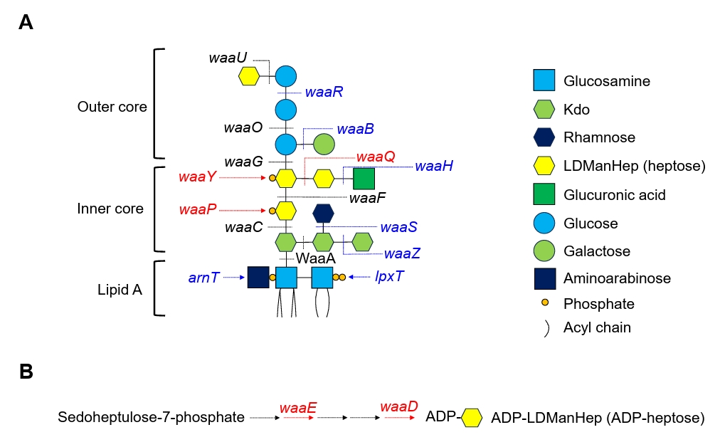
Fig. 2.Identification of deletion mutants exhibiting growth defects under envelope stress conditions. The indicated cells were serially diluted 10-fold from 108 to 104 cells/ml and spotted onto LB agar plates, with or without the indicated compounds (Van, vancomycin; EtOH, ethanol), or onto M9 minimal agar plates (M9) containing the indicated carbon sources (Glc, glucose; Lac, lactose; Gly, glycerol). No salt LB indicates LB agar plates without NaCl. The mutant strains with the defects in heptose biosynthesis or modification are shown in red color. Envelope stresses conditions that caused marked growth defects in the waaD and waaE mutants are also shown in red color. All data were obtained from two independent experiments.
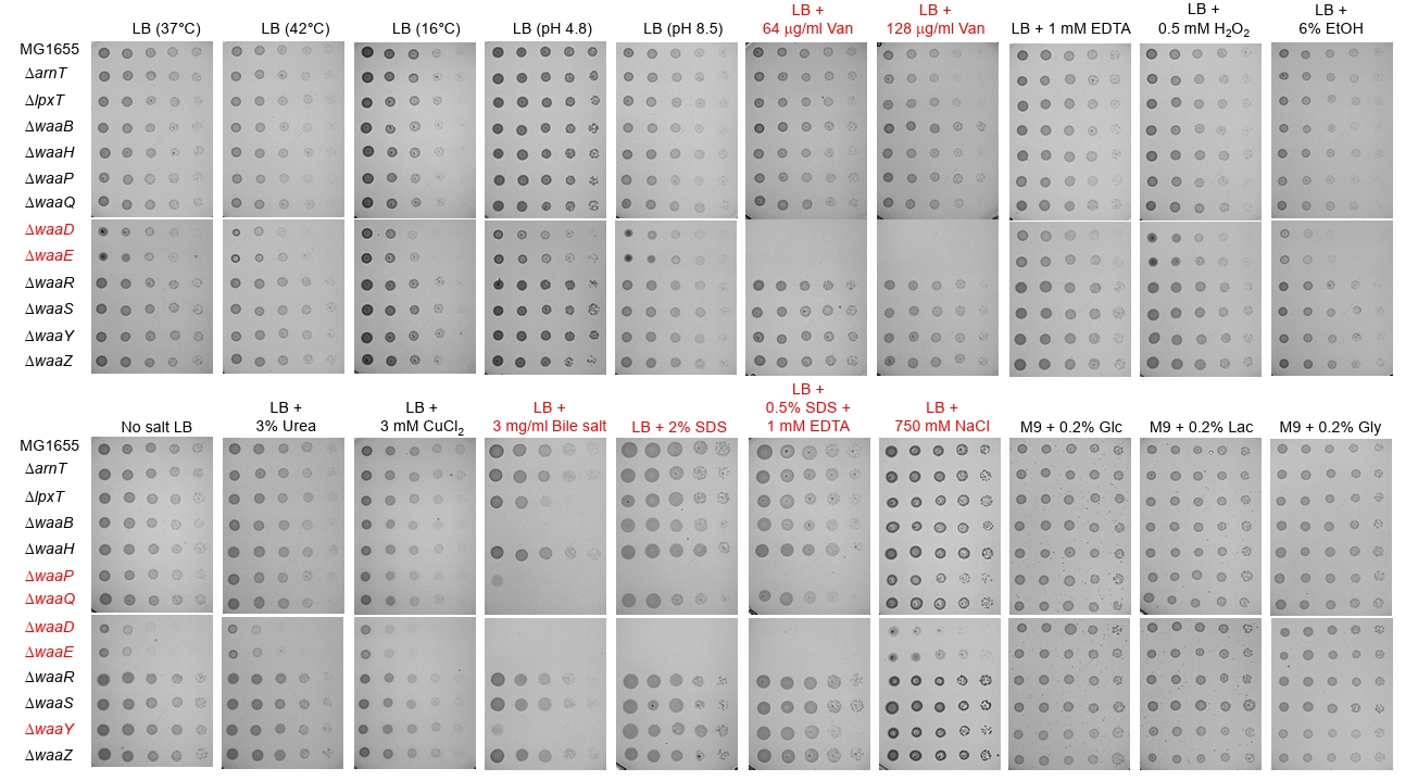
Fig. 3.The effects of deleting genes involved in heptose biosynthesis and modification on antibiotic susceptibility. The MIC values of the indicated antibiotics against the wild-type and indicated mutant strains were determined in MH medium. The relative MICs of the mutant strains were calculated using the MIC of the wild-type strain as the reference. The MICs of selected antibiotics are presented as colored bars. All data were obtained from two independent experiments.
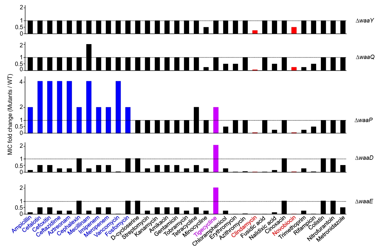
Fig. 4.Complementation of the phenotypes of the waaD and waaE mutants. (A) Complementation of growth defects of the waaD and waaE mutants under envelope stresses. The indicated cells were serially diluted 10-fold from 108 to 104 cells/ml and spotted onto an LB agar plate with or without the indicated compounds. No salt LB indicates LB agar plates without NaCl. (B) Complementation of the altered antibiotic susceptibility of the waaD and waaE mutants. The MIC of each antibiotic against the indicated strains was determined using 2-fold serial dilutions of the antibiotic. The final concentrations of the dilution change from 1,024 µg/ml to 0.125 μg/ml. All data were obtained from two independent experiments.
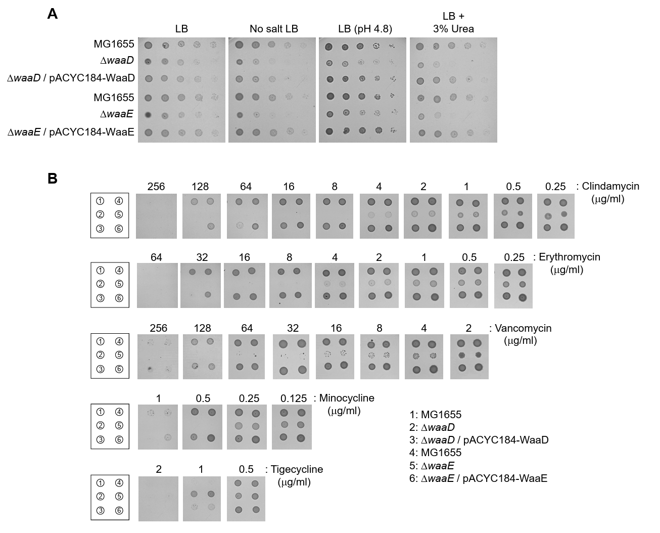
Fig. 5.Increased membrane permeability in the waaD and waaE mutants. (A) SDS-PAGE and silver staining analysis of LPS. LPSs extracted from the indicated strains were separated by SDS-PAGE and visualized by silver staining. (B) Assessment of membrane permeability using chlorophenyl red-β-D-galactopyranoside (CPRG). The indicated cells were cultured on LB agar plates containing 20 μg/ml CPRG for 20 h at 37°C. (C) Assessment of membrane permeability using the membrane-impermeable fluorescent dye SYTOX green. The indicated cells grown in LB medium to the exponential phase (OD600 = 1.0) were stained with SYTOX green (green) and then spotted onto a 1% agarose pad. Cells were visualized using a Nikon Eclipse Ni microscope. Scale bars = 20 μm. (D) The number of SYTOX green-positive cells among the cells imaged in panel (C). All data were obtained from two independent experiments.
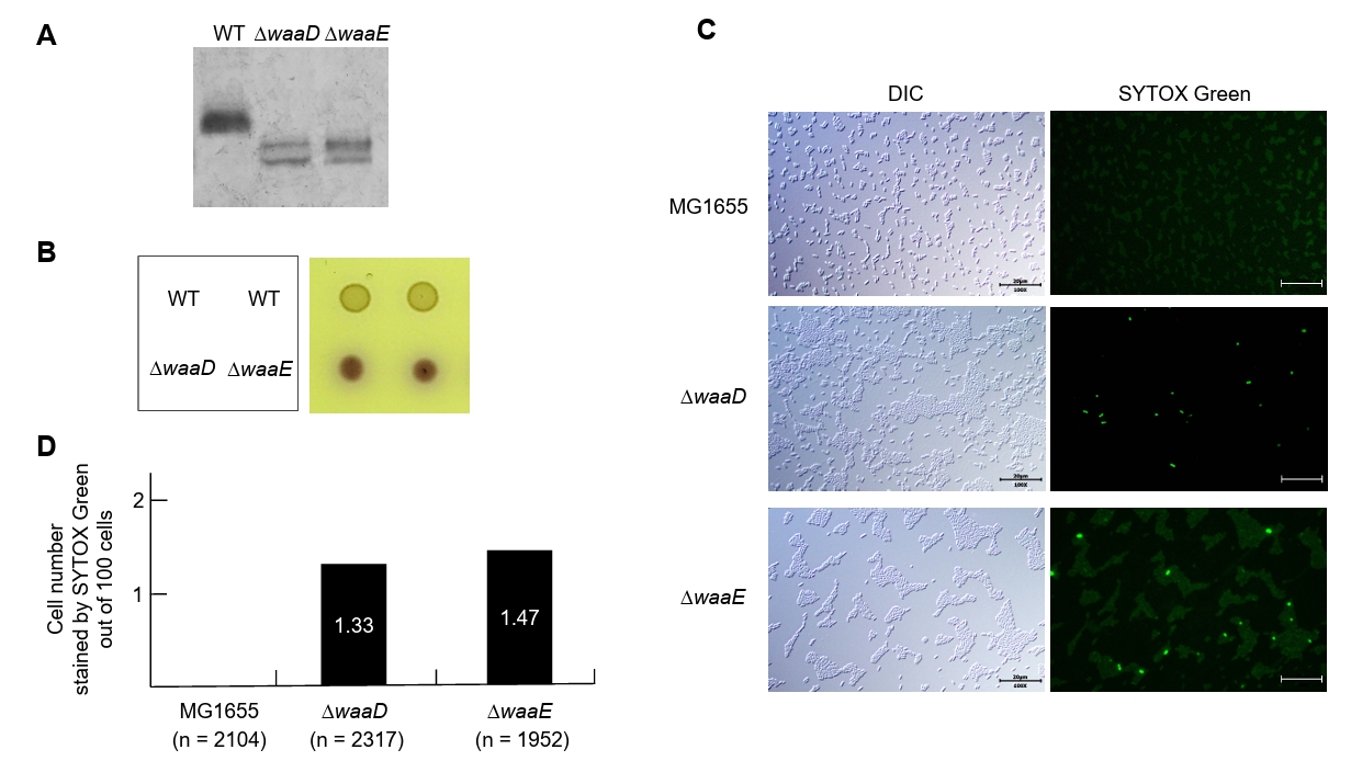
Fig. 6.The effect of putrescine biosynthesis on the phenotypes of the waaE mutant. (A) Isolation of suppressors of the waaE mutant. The cells of the indicated strains were serially diluted 10-fold from 108 to 104 cells/ml and spotted onto LB agar plates with or without clindamycin at the indicated concentrations. (B) Schematic representation of mini-Tn5 insertion sites. The mini-Tn5 insertion sites in suppressor mutants are indicated by red arrows. (C) The effect of putrescine biosynthesis on various phenotypes of the waaE mutant. The cells of the indicated strains were serially diluted 10-fold from 108 to 104 cells/ml and spotted onto LB agar plates with or without the indicated substances. (D) SDS-PAGE separation and silver staining analysis of LPS. LPSs extracted from the indicated strains were separated via SDS-PAGE and visualized by silver staining. All data were obtained from two independent experiments.
References
- Chen CW, Leimer N, Syroegin EA, Dunand C, Bulman ZP, et al. 2023. Structural insights into the mechanism of overcoming Erm-mediated resistance by macrolides acting together with hygromycin-A. Nat Commun. 14: 4196.ArticlePubMedPMCPDF
- Choi U, Son JE, Han G, Lee CR. 2025. Identification of genetic and environmental factors suppressing the lethality and antibiotic susceptibility mediated by depletion of LptD, a lipopolysaccharide transport protein. J Microbiol Biotechnol. 35: e2509011. ArticlePubMedPMC
- CLSI, Clinical and Laboratory Standards Institute. 2024. Performance standards for antimicrobial susceptibility testing, CLSI supplement M100, 34th edn. Clinical and Laboratory Standards Institute. PDF
- Datsenko KA, Wanner BL. 2000. One-step inactivation of chromosomal genes in Escherichia coli K-12 using PCR products. Proc Natl Acad Sci USA. 97: 6640–6645. ArticlePubMedPMC
- De Leon GP, Elowe NH, Koteva KP, Valvano MA, Wright GD. 2006. An in vitro screen of bacterial lipopolysaccharide biosynthetic enzymes identifies an inhibitor of ADP-heptose biosynthesis. Chem Biol. 13: 437–441. ArticlePubMed
- Desroy N, Moreau F, Briet S, Le Fralliec G, Floquet S, et al. 2009. Towards Gram-negative antivirulence drugs: New inhibitors of HldE kinase. Bioorg Med Chem. 17: 1276–1289. Article
- Di Lorenzo F, Duda KA, Lanzetta R, Silipo A, De Castro C, et al. 2022. A journey from structure to function of bacterial lipopolysaccharides. Chem Rev. 122: 15767–15821. ArticlePubMedLink
- Durka M, Tikad A, Perion R, Bosco M, Andaloussi M, et al. 2011. Systematic synthesis of inhibitors of the two first enzymes of the bacterial heptose biosynthetic pathway: Towards antivirulence molecules targeting lipopolysaccharide biosynthesis. Chemistry. 17: 11305–11313. ArticlePubMed
- Gevrekci AO. 2017. The roles of polyamines in microorganisms. World J Microbiol Biotechnol. 33: 204.ArticlePubMedPDF
- Gogry FA, Siddiqui MT, Sultan I, Haq QMR. 2021. Current update on intrinsic and acquired colistin resistance mechanisms in bacteria. Front Med (Lausanne). 8: 677720.ArticlePubMedPMC
- Huang SC, Panagiotidis CA, Canellakis ES. 1990. Transcriptional effects of polyamines on ribosomal proteins and on polyamine-synthesizing enzymes in Escherichia coli. Proc Natl Acad Sci USA. 87: 3464–3468. ArticlePubMedPMC
- Hummels KR. 2025. The regulation of lipid A biosynthesis. J Biol Chem. 301: 110556.ArticlePubMedPMC
- Kneidinger B, Marolda C, Graninger M, Zamyatina A, McArthur F, et al. 2002. Biosynthesis pathway of ADP-L-glycero-β-D-manno-heptose in Escherichia coli. J Bacteriol. 184: 363–369. ArticlePubMedPMC
- Langsrud S, Sundheim G. 1996. Flow cytometry for rapid assessment of viability after exposure to a quaternary ammonium compound. J Appl Bacteriol. 81: 411–418. ArticlePubMed
- Larsen RA, Wilson MM, Guss AM, Metcalf WW. 2002. Genetic analysis of pigment biosynthesis in Xanthobacter autotrophicus Py2 using a new, highly efficient transposon mutagenesis system that is functional in a wide variety of bacteria. Arch Microbiol. 178: 193–201. ArticlePubMedPDF
- Lee J, Park YH, Kim YR, Seok YJ, Lee CR. 2015. Dephosphorylated NPr is involved in an envelope stress response of Escherichia coli. Microbiology (Reading). 161: 1113–1123. ArticlePubMedPMC
- Lee HB, Park SH, Lee CR. 2021. The inner membrane protein LapB is required for adaptation to cold stress in an LpxC-independent manner. J Microbiol. 59: 666–674. ArticlePubMedPDF
- Lin J, Zhou D, Steitz TA, Polikanov YS, Gagnon MG. 2018. Ribosome-targeting antibiotics: Modes of action, mechanisms of resistance, and implications for drug design. Annu Rev Biochem. 87: 451–478. ArticlePubMedPMC
- Mitchell AM, Silhavy TJ. 2019. Envelope stress responses: Balancing damage repair and toxicity. Nat Rev Microbiol. 17: 417–428. ArticlePubMedPMCPDF
- Paradis-Bleau C, Kritikos G, Orlova K, Typas A, Bernhardt TG. 2014. A genome-wide screen for bacterial envelope biogenesis mutants identifies a novel factor involved in cell wall precursor metabolism. PLoS Genet. 10: e1004056. ArticlePubMedPMC
- Raetz CRH, Whitfield C. 2002. Lipopolysaccharide endotoxins. Annu Rev Biochem. 71: 635–700. ArticlePubMed
- Roth BL, Poot M, Yue ST, Millard PJ. 1997. Bacterial viability and antibiotic susceptibility testing with SYTOX green nucleic acid stain. Appl Environ Microbiol. 63: 2421–2431. ArticlePubMedPMCLink
- Ryu DB, Choi U, Han G, Lee CR. 2025. Inhibition of cardiolipin biosynthesis partially suppresses the sensitivity of an Escherichia coli mutant lacking OmpC to envelope stress. J Microbiol. 63: e2507004. ArticlePubMedPMCLink
- Son JE, Park SH, Choi U, Lee CR. 2024. Lytic transglycosylase repertoire diversity enables intrinsic antibiotic resistance and daughter cell separation in Escherichia coli under acidic stress. Antimicrob Agents Chemother. 68: e00372-24.ArticlePubMedPMCLink
- Sun J, Rutherford ST, Silhavy TJ, Huang KC. 2022. Physical properties of the bacterial outer membrane. Nat Rev Microbiol. 20: 236–248. ArticlePubMedPDF
- Tabor CW, Tabor H. 1985. Polyamines in microorganisms. Microbiol Rev. 49: 81–99. ArticlePubMedPMCLink
- VanOtterloo LM, Macias LA, Powers MJ, Brodbelt JS, Trent MS. 2024. Characterization of Acinetobacter baumannii core oligosaccharide synthesis reveals novel aspects of lipooligosaccharide assembly. mBio. 15: e03013-23.ArticlePubMedPMCLink
- Vester B, Douthwaite S. 2001. Macrolide resistance conferred by base substitutions in 23S rRNA. Antimicrob Agents Chemother. 45: 1–12. ArticlePubMedPMCLink
- Yethon JA, Heinrichs DE, Monteiro MA, Perry MB, Whitfield C. 1998. Involvement of waaY, waaQ, and waaP in the modification of Escherichia coli lipopolysaccharide and their role in the formation of a stable outer membrane. J Biol Chem. 273: 26310–26316. ArticlePubMed
- Yethon JA, Whitfield C. 2001. Purification and characterization of WaaP from Escherichia coli, a lipopolysaccharide kinase essential for outer membrane stability. J Biol Chem. 276: 5498–5504. ArticlePubMed
Citations
Citations to this article as recorded by

 MSK




 ePub Link
 Cite this Article
 XML Download