Skip Navigation
Skip to contents

Journal of Microbiology : Journal of Microbiology

OPEN ACCESS
SEARCH
Search

Articles

Page Path
HOME > J. Microbiol > Volume 64(10); 2026 > Review
Review
Epidemiology and spillover risk of SFTSV in animals across East Asia
Yun-Seon Shin1,2, Jeong Yoon Lee1,2, Jun-Gu Kang1,2,*orcid

DOI: https://doi.org/10.71150/jm.2608006
Published online: September 28, 2026

1Korea Zoonosis Research Institute, Jeonbuk National University, Iksan 54596, Republic of Korea

2Department of Veterinary and Animal Science, College of Veterinary Medicine, Jeonbuk National University, Iksan 54596, Republic of Korea

*Correspondence Jun-Gu Kang herculess@jbnu.ac.kr
• Received: August 4, 2026   • Accepted: August 26, 2026

© The Author(s), under exclusive licence to Microbiological Society of Korea 2026

This is an Open Access article distributed under the terms of the Creative Commons Attribution 4.0 International License (CC BY 4.0) (https://creativecommons.org/licenses/by/4.0/) which permits unrestricted use, distribution, and reproduction in any medium, provided the original work is properly cited.

  • 37 Views
  • 4 Download
  • Severe fever with thrombocytopenia syndrome (SFTS) is an emerging tick‑borne zoonosis in East Asia, yet the contribution of animals to virus maintenance and spillover remains incompletely characterized. This review examines current evidence of SFTS virus (SFTSV) infection across domestic livestock, companion animals, wildlife, and captive species, focusing on host range, geographic distribution, diagnostics, and reports of viral RNA and virus isolation. Available data indicate that SFTSV exposure and infection occur across a broad range of animal hosts, supporting a multi-host ecological framework rather than a simple single-reservoir interpretation. Domestic ruminants frequently exhibit high seroprevalence and may function as sentinels of enzootic activity, while occasional virus isolation from cattle demonstrates that viable virus can be recovered from naturally infected livestock, although this does not establish reservoir or amplification competence. Companion animals are considered a potential interface for interspecies transmission, as some studies have documented severe clinical disease, high viral loads, and cat-associated human transmission events supported by varying levels of epidemiological and genomic evidence. Studies in wildlife further document exposure and natural infection across diverse taxa, although their specific roles in viral maintenance remain unresolved. Interpretation of these findings depends heavily on the diagnostic modality: serology primarily indicates prior exposure, RNA detection supports current or recent infection, and virus isolation demonstrates that viable virus was present in the sampled material. Integrating animal infection data within an ecological framework reveals major knowledge gaps related to reservoir competence, infectious shedding, phylogenetic connections among animal, tick, and human cases, and implications for prevention. The review underscores the need for One Health surveillance that incorporates clinical data from companion animals and applies evidence‑based interpretation to improve recognition of animal–human interfaces associated with potential spillover risk.
Severe fever with thrombocytopenia syndrome (SFTS) is an emerging tick-borne infectious disease caused by severe fever with thrombocytopenia syndrome virus (SFTSV), currently classified as Bandavirus dabieense in the genus Bandavirus, family Phenuiviridae, and class Bunyaviricetes (ICTV, 2025). The first case of SFTS was reported in China in 2010 (Yu et al., 2011), followed by subsequent reports in the Republic of Korea (ROK), Japan, and Taiwan (Lee et al., 2024; Peng et al., 2020; Takahashi et al., 2014). Haemaphysalis longicornis is considered a principal vector of SFTSV and is widely distributed in East Asia, where SFTS is endemic, particularly in China, the ROK, and Japan (Casel et al., 2021; Kim and Park, 2023; Miao et al., 2020). The major clinical manifestations of SFTS include high fever, thrombocytopenia, leukopenia, and gastrointestinal and neurological symptoms (Casel et al., 2021; Seo et al., 2021).
SFTSV is a negative-sense single-stranded RNA (-ssRNA) virus with a tripartite genome consisting of the L (large), M (medium), and S (small) segments (Casel et al., 2021). The L segment encodes the RNA-dependent RNA polymerase (RdRp), which is essential for viral replication. The M segment encodes a glycoprotein precursor that is cleaved by host proteases into two envelope glycoproteins, Gn and Gc. Gn plays a critical role in receptor recognition and viral attachment (Ni et al., 2015), whereas Gc, a class II fusion protein, mediates membrane fusion and subsequent internalization (Halldorsson et al., 2016). These glycoproteins are key determinants of viral entry and major immunogenic targets for vaccine development. The S segment uses ambisense coding, encoding the nucleocapsid protein (NP) and the nonstructural protein (NSs). NP encapsidates viral RNA to form the ribonucleoprotein complex, whereas NSs suppresses host interferon signaling and functions as a major virulence factor (Casel et al., 2021; Hong et al., 2019; Khalil et al., 2021; Kitagawa et al., 2018; Zhou et al., 2013).
The case fatality rate (CFR) of SFTS varies among countries and has been reported as approximately 5.3% in China, 18.7% in the ROK, and 23% in Japan (Kim et al., 2023; Matsuu et al., 2019; Zhan et al., 2017). This regional variation has been discussed in relation to geographic and ecological factors, as well as genotype distribution (Casel et al., 2021). Currently, no effective antiviral agents or licensed vaccines are available for SFTSV infection. Although SFTSV is primarily transmitted by tick bites, human-to-human transmission via direct contact with blood, body fluids, or secretions of infected individuals has been documented. Animal-associated human infections, particularly those involving close contact with infected cats, have also been reported (Casel et al., 2021; Wu et al., 2022; Yamanaka et al., 2020). Accordingly, SFTS is regarded as an important zoonotic disease that requires close surveillance, not only because of vector-borne transmission but also because of potential infection through animal contact.
Although numerous epidemiological and clinical studies have focused on human infection, systematic investigations of SFTSV infection in animals and the ecological roles of different host species remain limited (Casel et al., 2021). Therefore, this review aims to summarize animal species with reported evidence of SFTSV exposure or infection, distinguish among serological, molecular, and virus-isolation evidence, and evaluate their implications without equating detection with reservoir or amplification competence.
Relevant studies were identified through searches of PubMed and Google Scholar using the terms “SFTS,” “SFTSV,” “animal,” and “virus isolation,” as well as combinations of these terms with specific host categories, including livestock, companion animals, wildlife, rodents, cats, and dogs. Studies published from database inception through January 2026 were included if they reported serological evidence, viral RNA detection, or virus isolation from naturally infected animals in East Asia and provided sufficient host-specific information, including host species, specimen type, diagnostic method, and numbers tested and positive, for data extraction.
Studies involving experimental infections, focusing exclusively on ticks or humans, or lacking identifiable information on host species, specimen type, diagnostic method, or the numbers of tested and positive animals were excluded from the aggregated analysis. Because study populations, specimen types, diagnostic assays, and sampling designs varied among studies, the combined data were interpreted as descriptive summaries rather than pooled prevalence estimates. In addition, serological and molecular tests were not consistently performed on, or reported for, the same individual animals; therefore, some animals may have been represented in more than one diagnostic category.
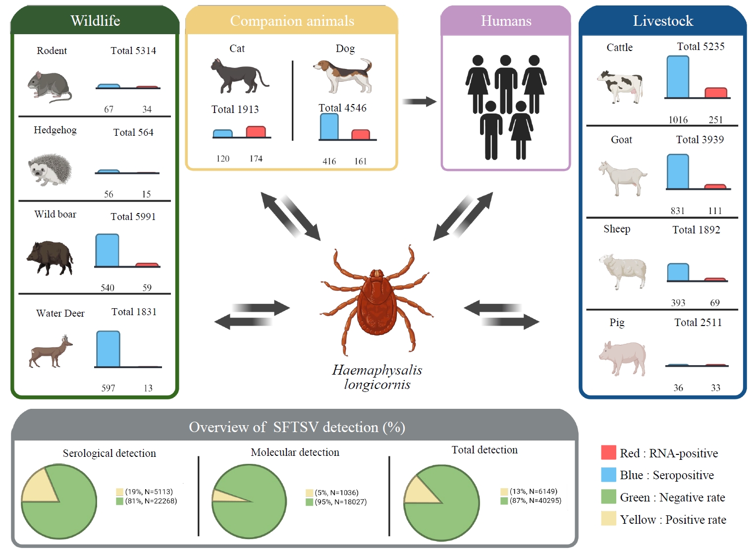
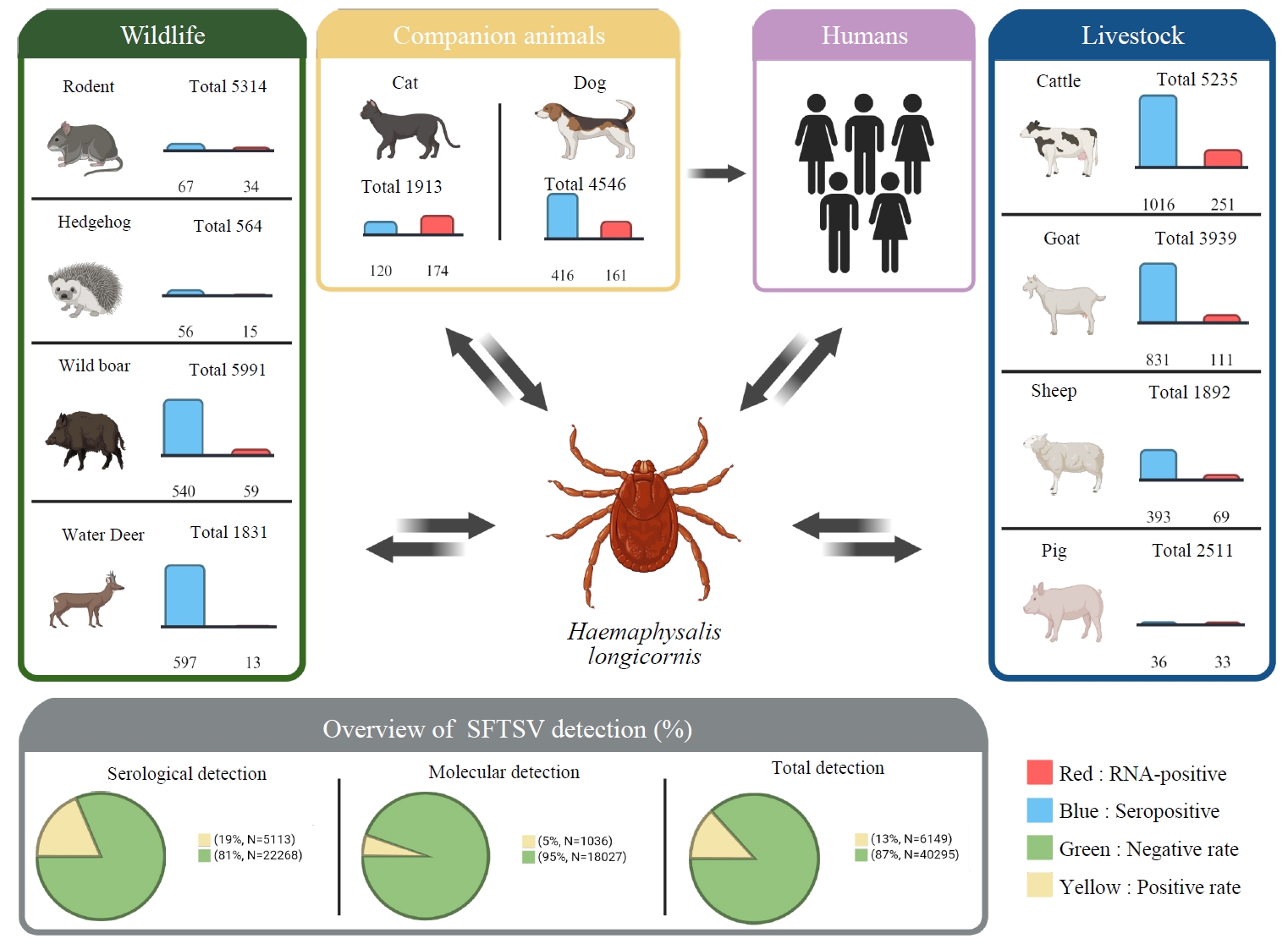
The animal exposure and infection data summarized in Table 1 indicate that SFTSV has been detected in a wide range of vertebrate hosts across East Asia. Figure 1, based on the data presented in Table 1, provides a schematic overview of SFTSV detection across major animal host groups. It also schematically illustrates potential connections among these hosts through shared tick vectors within the animal–tick–human transmission interface. For the purpose of synthesis, some host categories were grouped at higher taxonomic levels (e.g., “birds” or “rodents”), particularly where individual species were inconsistently reported or where numerous species were represented by limited sample sizes. This grouping was applied to facilitate comparative interpretation across studies. Across more than 40,000 aggregated animal observations reported in studies from multiple endemic countries, serological and molecular evidence have been reported in livestock, companion animals, wildlife, and captive species. Because serological and molecular testing were not consistently performed on the same individuals, this number should not be interpreted as the number of unique animals. The reported magnitude and pattern of detection vary substantially among host groups and study settings (Cheng et al., 2025; Li et al., 2016; Zhao et al., 2022). A key feature of these data is the heterogeneity of diagnostic profiles across species. Some host groups are characterized mainly by serological positivity, whereas others show more frequent RNA detection or occasional virus isolation. These contrasting patterns may reflect differences in exposure and infection dynamics, but they are also influenced by specimen type, diagnostic method, sampling time, study population, and sampling context.
Livestock
Domestic livestock, particularly cattle, goats, and sheep, are among the most extensively surveyed host groups in endemic regions. Across studies, these species frequently show relatively high seroprevalence, consistent with repeated exposure to SFTSV in tick-endemic agricultural environments (Ding et al., 2014; Kang et al., 2018a; Niu et al., 2013). In the aggregated dataset, cattle constitute one of the largest groups of tested animals, with substantial numbers of seropositive individuals, whereas goats and sheep show similar patterns of antibody detection. By contrast, RNA detection rates in livestock are generally lower than seroprevalence. This pattern, characterized by high antibody positivity and comparatively infrequent RNA detection, has been reported across multiple studies (Chae et al., 2024; Ding et al., 2014; Niu et al., 2013). These findings suggest that although exposure is common, detection of viral RNA at a single time point is less frequent in these species. Virus isolation has also been reported in livestock, including cattle, although such findings remain limited (Chae et al., 2024). Overall, livestock are characterized by frequent serological positivity and comparatively lower molecular positivity in cross-sectional sampling.
Companion animals
Companion animals, particularly cats and dogs, have been reported to show both serological and molecular evidence of SFTSV infection in endemic regions. In the available literature, RNA detection has been reported more frequently in companion animals than in livestock and some wildlife host groups, particularly among clinically affected individuals (Kang et al., 2019, 2020; Kuan et al., 2023b; Matsuu et al., 2019). In cats, RNA-positive cases have been repeatedly reported in both clinical and field studies, and relatively high viral loads have been observed in naturally infected individuals (Kang et al., 2020; Kuan et al., 2023b; Matsuu et al., 2019). However, this apparent difference should be interpreted cautiously because many companion-animal studies preferentially sampled clinically affected animals, which may have increased the likelihood of detecting viral RNA. In dogs, evidence of exposure, including antibodies and occasional detection of RNA, has also been reported (Ishijima et al., 2023; Kang et al., 2019). However, compared with cats, reports of molecular positivity and clinical severity vary among studies. Overall, companion animals show evidence of both prior exposure and natural infection, but the reported frequency of RNA detection should be interpreted in light of clinical sampling bias and differences in study design.
Wildlife
Wildlife represents the most taxonomically diverse host category in Table 1 and includes rodents, hedgehogs, wild boars, deer, and various carnivores (Hayasaka et al., 2016; Kaneko et al., 2023; Ni et al., 2015; Yu et al., 2019; Zhao et al., 2022). Evidence of SFTSV exposure and natural infection in wildlife includes serological and molecular findings, although patterns vary substantially among species. Rodents are among the most frequently studied wildlife hosts and have repeatedly been reported as RNA-positive across multiple geographic regions (Gu et al., 2022; Ni et al., 2015). In some studies, virus isolation has also been achieved from rodent species, confirming that a viable virus was present in some naturally infected animals without establishing a reservoir or maintenance competence. Hedgehogs have also been reported to be both seropositive and RNA-positive, and some studies have described virus isolation (Sun et al., 2017; Zhao et al., 2022). Although the number of studies is less than that for rodents, positive findings have been reported across multiple datasets. By contrast, larger mammals such as wild boars and deer are more often characterized by high seroprevalence with lower rates of RNA detection (Hayasaka et al., 2016; Yu et al., 2019). This pattern is similar to that observed in livestock. Other wildlife species, including carnivores, have been reported as seropositive or RNA-positive, but the available data remain limited (Kaneko et al., 2023).
Summary of host-associated patterns
Taken together, the data summarized in Table 1 show descriptive differences in reported detection profiles across host groups. Livestock and large wild mammals are typically characterized by high seroprevalence with relatively low reported RNA detection, whereas molecular evidence of infection has been reported more frequently in companion animals and some small wildlife species in the available literature. However, these differences should not be interpreted as directly comparable host-specific detection rates, as study designs and sampling contexts varied substantially. Virus isolation has been reported in a subset of hosts, including rodents, hedgehogs, companion animals, and cattle (Chae et al., 2024; Matsuu et al., 2019; Ni et al., 2015; Zhao et al., 2022). These patterns indicate that SFTSV exposure and natural infection have been documented across multiple host groups, with variation in detection type and frequency depending on species, study design, and sampling context (Cheng et al., 2025; Li et al., 2016; Zhao et al., 2022).
Interpretation of animal SFTSV data requires careful attention to the type of diagnostic evidence reported because serology, RNA detection, and virus isolation do not represent equivalent biological evidence of host infection or transmission potential. Although these methods are often presented together in animal surveillance studies, each captures a different dimension of host–virus interaction. Serological positivity indicates prior exposure, molecular detection supports current or recent infection by demonstrating the presence of viral nucleic acid at the time of sampling, and virus isolation provides direct evidence that a viable, replication-competent virus was present in the sampled material (Casel et al., 2021; Jiao et al., 2012; Kim and Park, 2023). Accordingly, interpretation of host-associated SFTSV data depends not only on whether a species is reported as positive, but also on how that positivity was established.
This distinction is particularly important in the context of the broad host range summarized in Table 1. The literature includes species with high seroprevalence but limited RNA detection, species with repeated molecular detection but less extensive serological support, and a smaller subset from which viable virus has been isolated following natural infection. These contrasting diagnostic profiles have different interpretive implications. Diagnostic modality should therefore be treated as a central component of interpretation rather than as a minor technical detail. However, none of these diagnostic findings alone establishes reservoir, amplification, or onward-transmission competence.
Serological evidence as an indicator of cumulative exposure
Serological assays have been the most widely used tools in animal SFTSV research, particularly in livestock and wildlife studies, because they are practical for large-scale field surveillance and useful for estimating cumulative exposure within host populations (Ding et al., 2014; Hayasaka et al., 2016; Kang et al., 2018a; Niu et al., 2013; Yu et al., 2019). Across endemic regions, Enzyme-linked immunosorbent assay (ELISA), Immunofluorescence assay (IFA), Virus neutralization test (VNT), Gaussia luciferase immunoprecipitation system (GLIPS), Focus reduction neutralization test (FRNT), Immunoperoxidase test (IPT), Single-Round Infectious Particles neutralization assay (SRIP neutralization assay), and related platforms have revealed substantial seropositivity in cattle, goats, sheep, deer, and wild boars, consistent with repeated exposure to SFTSV in tick-endemic environments (Ding et al., 2014; Hayasaka et al., 2016; Niu et al., 2013; Yu et al., 2019). The principal strength of serology is its ability to capture cumulative exposure within host populations. High seroprevalence across multiple studies is compatible with repeated exposure within local transmission environments, although the frequency and route of exposure cannot be determined from serology alone. This pattern is especially evident in domestic ruminants and large wild mammals, in which antibody prevalence is often substantial even when RNA detection is less frequent (Ding et al., 2014; Niu et al., 2013). In this sense, serological surveys are useful for identifying areas of enzootic circulation and recognizing host populations with substantial cumulative exposure. At the same time, seropositivity has clear interpretive limits. Antibody detection cannot distinguish recent from remote infection, cannot determine whether the host was viremic at the time of sampling, and does not establish whether infectious virus remains present. In cross-sectional studies, antibody-positive animals may have cleared infection long before sampling occurred (Ding et al., 2014; Niu et al., 2013). For that reason, serological evidence is best interpreted as evidence of prior exposure rather than proof of current infectiousness. This limitation becomes especially important when discussing reservoir hosts. High seroprevalence indicates repeated exposure within areas of viral circulation but does not define the functional role of a species or demonstrate reservoir competence. Demonstrating reservoir or amplification competence would require evidence that the host sustains viral replication at levels and for a duration sufficient to support onward transmission. Serological evidence alone cannot establish that point (Cheng et al., 2025; Li et al., 2016; Zhao et al., 2022). Accordingly, species supported only by antibody data are more appropriately described as exposed hosts than as confirmed reservoirs.
Molecular detection as evidence of current or recent infection
Compared with serology, detection of SFTSV RNA by reverse transcription polymerase chain reaction (RT-PCR), qRT-PCR, nested RT-PCR, or related assays demonstrates the presence of viral RNA in the tested specimen at the time of sampling and therefore supports current or recent infection (Chae et al., 2024; Gu et al., 2022; Kang et al., 2019, 2020; Matsuu et al., 2019). In the studies summarized in Table 1, RNA detection has been particularly important for documenting current or recent infection in host groups including cats, dogs, rodents, and cattle (Chae et al., 2024; Gu et al., 2022; Kang et al., 2019, 2020; Matsuu et al., 2019). However, RNA positivity is not equivalent to infectiousness. Detection of viral genetic material does not necessarily mean that an intact, viable virus is still present. Viral RNA may remain detectable after the infectious virus is no longer recoverable, particularly when sampling occurs late in infection or under conditions in which nucleic acid persistence is longer than the persistence of viable virus (Casel et al., 2021; Jiao et al., 2012; Kim and Park, 2023). Thus, molecular positivity provides stronger support than serology for current or recent infection, but does not provide the same evidence of viable virus as successful virus isolation. Interpretation of RNA detection is also strongly influenced by specimen type. As shown in Table 1, animal studies have used serum, plasma, whole blood, spleen, visceral tissues, and, in some companion animal studies, oral, ocular, or rectal specimens. These differences matter because the probability of detection depends on tissue distribution, viral burden, and timing of collection. Whole blood may capture viremia not detected in serum, whereas tissue-based sampling may yield higher positivity in necropsy-based wildlife studies. In companion animals, detection in non-blood specimens may indicate the presence of viral RNA in specimens that are potentially associated with shedding, although it does not, by itself, demonstrate infectious shedding (Ando et al., 2021; Kang et al., 2020; Matsuu et al., 2019). Reported RNA positivity should therefore not be interpreted as directly comparable across studies without accounting for specimen type, collection timing, and sampling context.
Timing of sampling is a second major determinant. Because many animal studies are cross-sectional, they capture only a narrow window of the infection course. If viremia is brief, naturally infected animals may be antibody-positive but PCR-negative at the time of sampling. This helps explain the recurrent pattern observed in livestock and some wildlife hosts: relatively high seroprevalence accompanied by lower RNA detection (Chae et al., 2024; Ding et al., 2014; Kang et al., 2018a; Niu et al., 2013). Similar profiles have been reported in wild boars and deer (Hayasaka et al., 2016; Yu et al., 2019). Such discrepancies do not necessarily indicate limited epidemiological relevance; they may instead reflect the transient window of RNA detectability. This issue is particularly useful for interpreting host-group differences. In cattle, goats, and sheep, high seroprevalence with comparatively infrequent RNA positivity may be consistent with repeated exposure and a limited period during which viral RNA is detectable (Chae et al., 2024; Ding et al., 2014; Kang et al., 2018a; Niu et al., 2013). By contrast, in companion animals such as cats, RNA detection is more often captured in clinically affected individuals, although this pattern may partly reflect the preferential sampling of clinically affected animals (Ando et al., 2021; Kang et al., 2020; Matsuu et al., 2019).
Virus isolation as evidence of a viable virus
Among the available diagnostic approaches, virus isolation provides the strongest biological evidence that viable, replication-competent SFTSV was present in the sampled material and could be recovered under laboratory conditions, beyond evidence of prior exposure or viral nucleic acid alone (Casel et al., 2021; Kim and Park, 2023; Yishuang et al., 2021). However, virus isolation alone does not establish that the sampled animal transmitted the virus under natural conditions or that the host species possesses reservoir or amplification competence. This distinction is central when considering the potential epidemiological role of an infected animal.
Isolation from Apodemus agrarius and hedgehogs demonstrates that viable SFTSV was present in some naturally infected animals. These findings identify these species as candidates for further ecological investigation but do not establish competence in maintenance or amplification. Demonstrating such roles would require evidence of sustained viremia, efficient host-to-tick transmission, and onward transmission under natural conditions.
Isolation from domestic cats is particularly notable because it is supported by a broader evidentiary pattern that includes repeated RNA detection, severe clinical disease, and cat-associated human transmission events. In one nosocomial outbreak, viable SFTSV was isolated from an infected cat and two veterinary personnel, and the complete S, M, and L genome segments showed 100% nucleotide identity, providing strong evidence of direct cat-to-human transmission (Yamanaka et al., 2020). However, even in this strongly supported event, the specific route of exposure—whether through blood, other body fluids, mucosal contact, droplets, or aerosols—was not determined. In this context, isolation is not merely a laboratory finding but part of a broader evidentiary pattern indicating that clinically affected cats may represent a relevant interface for potential human exposure during close contact with blood or body fluids. Isolation from cattle is also significant because it extends the available evidence beyond serological exposure alone. Recovery of viable virus from naturally infected cattle demonstrates that viable SFTSV can occasionally be recovered from livestock under field conditions (Chae et al., 2024). However, this finding remains insufficient to classify cattle or other livestock as reservoir or amplifying hosts.
At the same time, negative isolation findings must be interpreted cautiously. Virus recovery is technically demanding and depends on viral load, specimen type and quality, timing of collection, preservation conditions, and cell-culture methods. Failure to isolate SFTSV, therefore, does not necessarily demonstrate that a viable virus was absent or that the host lacks epidemiological relevance (Casel et al., 2021; Kim and Park, 2023). For that reason, positive isolation should be treated as strong evidence that a viable virus was present in the sampled material, whereas the absence of isolation should not be taken as proof of epidemiological irrelevance. Overall, successful isolation provides strong evidence that viable virus was present in the sampled material, but its ecological significance must be evaluated together with infection duration, shedding, host-to-tick transmission, contact patterns, and evidence of natural onward transmission.
Diagnostic heterogeneity and its implications for interpretation
A major challenge in synthesizing the animal SFTSV literature is the marked heterogeneity in study design and diagnostic strategy across host species, countries, and sampling settings. This heterogeneity affects not only reported positivity estimates but also the apparent relative importance of host groups. Some studies rely solely on serology, others combine serology with PCR, and only a minority attempt virus isolation. Assay platforms also vary substantially, including ELISA, IFA, VNT, RT‑PCR, nested RT‑PCR, and qRT‑PCR (Huang et al., 2019; Yishuang et al., 2021). Specimen heterogeneity further complicates interpretation. Some studies use serum only, whereas others analyze blood, plasma, spleen, viscera, or multiple specimen types. These differences are not trivial, as a PCR-negative result from one specimen type may not exclude a positive result from another. Companion animal studies that include non-blood specimens may detect viral RNA in oral, ocular, or rectal samples that serum-based surveys would not assess. However, RNA detection in these specimens does not by itself demonstrate infectious shedding (Ando et al., 2021; Matsuu et al., 2019). As a result, direct comparison of RNA detection rates across species is often difficult unless sampling frameworks are closely aligned. Clinical sampling context is another important source of variation. Many livestock and wildlife studies are based on apparently healthy field-caught or farmed animals, whereas some companion animal studies preferentially include clinically ill animals presented to veterinary settings. This difference can substantially influence the likelihood of RNA detection or virus isolation. Some of the apparent contrast between companion animals and other host groups may therefore reflect differences in sampling context as well as possible biological differences in infection dynamics. Geographic and ecological variation adds a final layer of complexity. Host density, habitat structure, agricultural practices, and local vector activity all influence the probability of exposure and detection (Cheng et al., 2025; Li et al., 2016). Because serological and molecular testing was not consistently performed on or reported for the same individual animals, the aggregated sample sizes in Table 1 may include overlap across diagnostic categories. The totals should therefore be interpreted as aggregated observations rather than counts of unique animals, independent denominators, or pooled sample sizes. Taken together, diagnostic results should be interpreted within their methodological and ecological settings rather than treated as directly interchangeable across studies.
An evidence-weighted framework for interpretation
Taken together, the available data support an evidence-weighted framework for interpreting animal SFTSV findings. Serology, RNA detection, and virus isolation provide complementary but non-equivalent evidence of exposure, current or recent infection, and the presence of viable virus, respectively; none alone establishes natural transmission, reservoir competence, or amplification competence (Casel et al., 2021; Kim and Park, 2023; Yishuang et al., 2021). This evidence-weighted approach helps avoid two common interpretive errors. The first is overinterpretation of serology, in which antibody-positive species are prematurely described as reservoirs or major transmission hosts despite lacking evidence of active or infectious virus. The second is the underinterpretation of stronger virological evidence, particularly when repeated RNA detection or virus isolation is treated as no more informative than serological exposure. A balanced interpretation is to describe serology-only species as exposed hosts, species with repeated molecular detection as hosts with evidence of current or recent infection, and isolation-positive species as hosts in which viable virus has been documented (Chae et al., 2024; Cheng et al., 2025; Zhao et al., 2022). These categories are complementary and should be interpreted together. The key question is not simply whether a host species has tested positive, but what type of positivity was observed, in which specimen, under what sampling conditions, and with what degree of biological confidence. Only by applying this layered interpretive approach can animal infection data be used to formulate cautious hypotheses regarding maintenance, amplification, and spillover, while recognizing that these hypotheses require additional ecological and transmission evidence across the animal–tick–human interface.
The epidemiological significance of animal SFTSV infection extends beyond vector-borne ecology alone. Although infected ticks remain the principal route of transmission in nature, available evidence indicates that direct exposure to infected blood or body fluids may result in transmission under certain circumstances, particularly at the animal–human interface (Hu et al., 2020; Yamanaka et al., 2020). The public health relevance of an infected animal, therefore, depends not only on its infection status but also on the opportunities for exposure created during handling, clinical care, restraint, necropsy, or contact with blood and other body fluids. Transmission implications should accordingly be interpreted in relation to host infection status, contact intensity, clinical context, and the evidentiary strength supporting non-vector spread (Kida et al., 2019; Kim and Park, 2023; Yamanaka et al., 2020).
Animal-to-human transmission
Reports of animal-to-human transmission of varying evidentiary strength have primarily involved companion animals, particularly cats. The evidentiary strength of reported events was assessed using epidemiological linkage, exclusion of alternative tick exposures, viral sequence identity, virus isolation, and availability of whole-genome comparisons. Case reports and outbreak investigations indicate that humans may be exposed to SFTSV through close contact with infected cats in veterinary settings, where contact with blood, saliva, respiratory secretions, or other body fluids may occur, although the specific infectious material responsible for transmission has not been identified (Ando et al., 2021; Kida et al., 2019; Yamanaka et al., 2020). These findings are epidemiologically relevant because they demonstrate the potential for zoonotic exposure in real clinical settings. The public health implications of feline infection have been further discussed in light of reports describing severe systemic disease and relatively high viral loads in naturally infected cats (Ando et al., 2021; Matsuu et al., 2019; Osako et al., 2024). Under such conditions, bites, scratches, mucosal exposure, and direct contact with blood or body fluids that may contain the virus have been proposed as possible routes; however, strongly supported transmission events have also occurred in the absence of recognized bites or scratches. Accordingly, clinically affected cats may serve as a relevant interface for interspecies exposure, as close contact with humans often occurs during veterinary care (Ando et al., 2021; Kida et al., 2019; Yamanaka et al., 2020). The reported cat-associated events differ in evidentiary strength. One report described a veterinarian who developed SFTS after caring for and performing autopsies on infected cats; an identical 421-nucleotide viral sequence was identified in the patient and two cats, although the specific source cat, transmission route, and whole-genome linkage were not established (Kida et al., 2019). By contrast, a nosocomial outbreak involving an infected cat and two veterinary personnel provided stronger virological evidence: viable virus was isolated from all three individuals, and the complete S, M, and L genome segments showed 100% nucleotide identity (Yamanaka et al., 2020). This outbreak therefore provides stronger evidence of direct cat-to-human transmission, although the precise route of exposure remains undetermined. Dogs have also been associated with human exposure under certain circumstances, although evidence for direct dog-to-human transmission remains limited and is less extensive than that for cats. Naturally infected dogs have been reported as RNA-positive and clinically affected in endemic regions (Kang et al., 2019; Nam et al., 2020), and a human infection temporally associated with close contact during tick removal has also been described (Chung et al., 2020). However, exposure to the infected tick cannot be clearly distinguished from direct transmission from the dog in this setting. While these findings do not support classifying dogs at the same level as cats in terms of evidence for zoonotic spillover, they suggest that dogs may be associated with human exposure in specific contexts, particularly during tick removal or close handling of clinically affected animals (Chung et al., 2020; Kang et al., 2019; Nam et al., 2020). Taken together, zoonotic transmission risk appears to depend not only on host susceptibility but also on patterns of contact, route of exposure, and environmental context. Companion animals live in settings characterized by close human contact, and in some situations, infections in these hosts may more readily translate into human exposure. These considerations highlight the importance of including companion animals in assessments of interspecies transmission risk, while distinguishing events by their evidentiary strength (Ando et al., 2021; Kida et al., 2019; Yamanaka et al., 2020).
Animal-to-animal transmission
Compared with animal-to-human transmission, direct evidence for animal-to-animal transmission remains limited. Nevertheless, non-vector animal-to-animal transmission is biologically plausible under certain conditions (Chae et al., 2024; Matsuu et al., 2019; Zhao et al., 2022). Recovery of viable virus from naturally infected animals indicates that replication-competent SFTSV was present in the sampled material, suggesting that close contact among animals could permit non-vector transmission under appropriate conditions (Chae et al., 2024; Matsuu et al., 2019; Zhao et al., 2022). However, virus isolation alone does not demonstrate direct animal-to-animal transmission. This possibility may be particularly relevant in companion animal settings. Infected cats can develop severe disease accompanied by high viral burdens, creating conditions in which contact with blood, saliva, ocular discharge, or other body fluids may plausibly expose other animals in shelters, veterinary hospitals, or multi-animal households (Ando et al., 2021; Matsuu et al., 2019; Osako et al., 2024). The reported cat-to-human events, including the genomically linked outbreak, do not demonstrate direct cat-to-cat transmission, and no equivalent genomic linkage between naturally infected animals has been established in these reports. However, current field evidence remains insufficient to determine how often such direct transmission occurs or whether it contributes meaningfully to sustained transmission outside periods of tick activity. A similar uncertainty applies to wildlife. The recovery of viable virus from wildlife hosts such as rodents or hedgehogs confirms natural infection but does not establish non-vector animal-to-animal transmission (Ni et al., 2015; Zhao et al., 2022). Moreover, most field studies cannot distinguish direct transmission from shared exposure to infected ticks. As a result, animal-to-animal spread remains a plausible but incompletely defined component of SFTSV transmission ecology rather than a firmly established maintenance route.
Non-vector transmission and occupational implications
Available evidence indicates that tick-borne transmission remains the dominant natural pathway of SFTSV circulation (Hu et al., 2020; Zhuang et al., 2018). Nevertheless, documented person-to-person transmission through exposure to infected blood demonstrates that contact-based transmission is biologically possible and provides a relevant framework for interpreting animal-associated exposure (Wu et al., 2022). The precise route involved in individual animal-associated events often remains uncertain, and the relative contributions of blood, other body fluids, mucosal exposure, droplets, or aerosols have not been fully resolved.
The possibility of non-vector transmission is particularly relevant in clinical and occupational settings. Clinically affected animals with high viral loads may create opportunities for exposure during examination, treatment, restraint, specimen collection, necropsy, or handling of materials potentially contaminated with blood or body fluids. Veterinarians, veterinary nurses, shelter workers, farmers, wildlife handlers, and pet owners may therefore encounter blood or other body fluids that may contain viral RNA and, in some reported cases, viable virus (Ando et al., 2021; Chung et al., 2020; Kida et al., 2019; Yamanaka et al., 2020).
Prevention strategies should therefore address both tick-borne transmission and possible contact-based exposure. In settings involving clinically ill animals, appropriate personal protective equipment, careful handling of blood and body fluids, safe specimen collection, and precautions during necropsy are warranted (Ando et al., 2021; Kida et al., 2019; Yamanaka et al., 2020). These measures are particularly relevant when the infection status of an animal is unknown but clinical and epidemiological features are compatible with SFTSV infection.
Overall, non-vector transmission is less frequently documented than tick-borne transmission, but its true frequency, specific routes, and broader epidemiological contribution remain uncertain. This uncertainty emphasizes the need for improved infection-control awareness and systematic investigation of occupational and veterinary-associated exposures.
The broad host range and heterogeneous infection profiles summarized in this review are consistent with SFTSV circulation within a multi-host ecological system rather than a simple single-reservoir structure (Cheng et al., 2025; Li et al., 2016; Zhao et al., 2022). Across East Asia, SFTSV exposure and natural infection have been documented in livestock, companion animals, and diverse wildlife species, although the type and strength of evidence differ substantially among host groups. Collectively, these findings suggest a transmission network in which host roles vary with ecological setting, vector activity, host abundance, and opportunities for human contact (Cheng et al., 2025; Hu et al., 2020; Zhuang et al., 2018). This interpretation shifts attention away from the search for a single definitive reservoir and toward interactions among hosts, ticks, and human exposure settings. Current evidence is more consistent with a framework in which different vertebrate hosts occupy different positions within the broader transmission system (Cheng et al., 2025; Li et al., 2016; Zhao et al., 2022). Within this framework, livestock may be particularly useful as indicators of enzootic exposure. Domestic ruminants repeatedly show substantial serological evidence of exposure in endemic agricultural settings (Ding et al., 2014; Niu et al., 2013), making them useful for identifying areas with substantial enzootic exposure. Wildlife may contribute to broader ecological circulation, although the extent and nature of this contribution remain species- and context-dependent (Cheng et al., 2025; Li et al., 2016; Zhao et al., 2022). Companion animals occupy a different position: their clearest documented significance lies at a high-contact interface where animal infection may become directly relevant to human exposure (Ando et al., 2021; Kida et al., 2019; Yamanaka et al., 2020). These distinctions have direct implications for surveillance. If SFTSV circulation is understood within a multi-host framework, surveillance limited to human cases or infected ticks will capture only part of the transmission landscape. Human case surveillance identifies recognized disease, and tick surveillance reflects vector infection pressure, whereas animal surveillance provides complementary information on host exposure, current or recent infection, and potential spillover-relevant interfaces (Cheng et al., 2025; Kuan et al., 2023a; Li et al., 2016). Different animal sectors may therefore serve different surveillance purposes: livestock serology may be useful for mapping enzootic activity, whereas molecular detection or virus isolation in companion animals and selected wildlife hosts may be more informative for identifying host groups or settings in which current infection or viable virus has been documented (Chae et al., 2024; Zhao et al., 2022). Taken together, these patterns reinforce the need for a One Health framework integrating wildlife ecology, veterinary medicine, livestock management, vector biology, and human public health (Casel et al., 2021; Kim and Park, 2023). Spillover risk may emerge from the overlap of these domains rather than from any one domain alone: infected ticks feed on multiple vertebrate hosts, infected animals may, under some circumstances, create opportunities for direct exposure, and local ecological conditions shape how often animals, vectors, and humans come into contact (Hu et al., 2020; Kida et al., 2019; Yamanaka et al., 2020). For this reason, preparedness is likely to be most effective when animal surveillance, tick monitoring, veterinary awareness, and human public health responses are treated as connected rather than separate systems (Casel et al., 2021; Cheng et al., 2025; Kim and Park, 2023). From a practical perspective, this integrated approach is especially relevant in farms, veterinary clinics, animal shelters, and wildlife management settings, where animal infection may function both as an ecological signal and as a potential occupational concern (Ando et al., 2021; Chung et al., 2020; Kida et al., 2019; Yamanaka et al., 2020). In such environments, an infected animal may not only indicate local circulation of SFTSV but, in some circumstances, may also identify a setting in which spillover-relevant exposure could occur.
Overall, SFTSV ecology is best interpreted through a host–vector–human framework in which different animal groups occupy different positions in ecological circulation and spillover-relevant interfaces (Cheng et al., 2025; Li et al., 2016; Zhao et al., 2022). One Health surveillance should therefore focus not only on infected species, but also on the interfaces that connect enzootic circulation with human exposure risk (Casel et al., 2021; Cheng et al., 2025; Kim and Park, 2023).
Despite the growing literature on animal SFTSV infection, major uncertainties remain regarding which host species may contribute to viral maintenance, which primarily reflect exposure, and which may be most relevant to spillover into humans (Cheng et al., 2025; Li et al., 2016; Zhao et al., 2022). Current studies have established that a wide range of animals can be seropositive, RNA-positive, and in some cases, yield viable virus upon isolation. However, these findings do not by themselves identify the hosts that matter most epidemiologically. In other words, the field has advanced substantially in documenting host breadth, but it still lacks comparable resolution in defining host function.
One of the most important unresolved issues is reservoir competence. Evidence of exposure or infection does not necessarily indicate that a host can efficiently infect feeding ticks, sustain transmission over time, or consistently contribute to local maintenance cycles (Cheng et al., 2025; Li et al., 2016). This issue is especially relevant for wildlife species that are often discussed as putative reservoirs on the basis of repeated field detection alone. Rodents remain plausible candidates because of their abundance, wide distribution, and repeated molecular detection (Gu et al., 2022; Ni et al., 2015), whereas hedgehogs have attracted particular attention because repeated field detection and virus isolation identify them as candidates for further investigation of their potential amplification roles (Zhao et al., 2022). However, direct evidence of efficient onward transmission remains insufficient, and for most wildlife hosts, current evidence does not clarify whether, or how consistently, such roles operate across ecological settings.
A second major gap concerns duration of viremia, shedding of viable virus, and host infectiousness. Most animal studies are cross-sectional and therefore identify whether infection has occurred, but not how long infected animals remain viremic, have detectable viral RNA, shed viable virus, or create opportunities for transmission (Ando et al., 2021; Chae et al., 2024; Matsuu et al., 2019). This limitation is particularly important at the animal–human interface. The public health relevance of an infected animal depends not only on whether the virus is detectable at a single time point, but also on whether viable virus is present, in what quantity, in which specimens or body fluids, and for how long it remains detectable or recoverable. Without longitudinal data, estimates of host-specific windows of potential infectiousness remain uncertain.
A third unresolved issue is comparative host importance. The available literature suggests that infected hosts are not epidemiologically equivalent, yet direct comparative data remain scarce (Chae et al., 2024; Cheng et al., 2025; Zhao et al., 2022). Some host groups appear to be characterized mainly by repeated exposure, whereas others show stronger evidence supporting current or recent infection or potential spillover relevance. However, these differences are still inferred largely from separate studies that vary in design, specimen type, and sampling context. As a result, it remains difficult to reliably compare host species in terms of their potential contributions to maintenance, amplification, or spillover.
Another major gap concerns phylogenetic linkage across hosts, ticks, and humans. Although several studies have reported molecular detection and sequence analysis in animals (Lee et al., 2017), integrated datasets directly connecting viral sequences across wildlife, livestock, companion animals, ticks, and human cases remain limited. Although whole-genome identity has been demonstrated in a small number of veterinary-associated cat-to-human events, comparable genomic linkage remains unavailable for most animal-associated infections and for most proposed animal-to-animal or host-to-tick transmission pathways. This restricts inference about transmission direction, bridge hosts, and the extent to which infections detected in different sectors belong to the same local transmission cycle (Cheng et al., 2025; Li et al., 2016). More comprehensive cross-sector phylogenetic analyses would substantially improve the interpretation of how enzootic circulation translates into human exposure or disease risk. Methodological heterogeneity further complicates the field. Animal studies vary widely in serological platforms, molecular assays, specimen types, host clinical status, and sampling designs (Huang et al., 2019; Matsuu et al., 2021; Yishuang et al., 2021). These differences weaken comparability across host species, countries, and study settings and may partly explain apparent variation in reported positivity or host importance. Some observed contrasts are likely biological, but others may reflect methodological inconsistency rather than underlying ecological differences. Greater standardization in sampling and diagnostic strategy is therefore essential if future studies are to support stronger comparative inference.
Taken together, these gaps limit the extent to which current animal infection data can be translated into precise surveillance priorities and prediction of spillover risk. Future work should move beyond descriptive host lists and focus more directly on reservoir competence, longitudinal shedding dynamics, comparative host infectiousness, and integrated phylogenetic linkage across the animal–tick–human interface (Cheng et al., 2025; Li et al., 2016; Zhao et al., 2022). Progress in these areas will be necessary to transform the current literature from a catalog of host detections into a mechanistic understanding of how SFTSV persists and spills over in real ecological systems.
The evidence reviewed here indicates that SFTSV circulates across a wide range of animal hosts in East Asia and is more appropriately understood within a multi-host ecological system rather than a single-reservoir framework (Cheng et al., 2025; Li et al., 2016). Although animal exposure and infection are widely reported, their epidemiological significance varies according to the type of diagnostic evidence. Serological findings primarily reflect prior exposure, molecular detection supports current or recent infection, and virus isolation demonstrates that viable virus was present in the sampled material. However, none of these diagnostic findings alone establishes reservoir, amplification, or onward-transmission competence. Companion animals, particularly cats, occupy a relatively important position because natural infection, clinical disease, virus isolation, and genomically supported cat-associated human transmission have all been documented (Ando et al., 2021; Kida et al., 2019; Matsuu et al., 2019; Yamanaka et al., 2020). In contrast, livestock often reflect enzootic exposure at the landscape level, whereas findings in wildlife document broad exposure and natural infection but do not yet define species-specific maintenance roles (Chae et al., 2024; Cheng et al., 2025; Ni et al., 2015; Zhao et al., 2022).
Taken together, these findings highlight three key points. First, host roles should be interpreted within a multi-host ecological framework rather than assigned to a single presumed reservoir. Second, host importance must be evaluated using an evidence-weighted approach that distinguishes prior exposure from current or recent infection and from the presence of viable virus. Third, spillover risk is shaped not only by host infection status but also by the position of that host within the animal–tick–human interface (Cheng et al., 2025; Kim and Park, 2023; Li et al., 2016). Accordingly, effective preparedness requires a One Health approach that integrates animal surveillance, veterinary recognition, vector monitoring, and human public health responses (Cheng et al., 2025; Kim and Park, 2023; Li et al., 2016). Future research should move beyond descriptive reports of host detection and focus more directly on reservoir competence, infectiousness, shedding dynamics, and cross-sector transmission linkages. Addressing these questions will be essential for identifying the hosts and interfaces that may contribute most meaningfully to viral maintenance and spillover in East Asia (Cheng et al., 2025; Li et al., 2016; Zhao et al., 2022).
Fig. 1.
Overview of SFTSV detection across animal hosts and the animal–tick–human interface. This figure summarizes the reported serological and molecular detection of severe fever with thrombocytopenia syndrome virus (SFTSV) across major animal host groups, including wildlife, companion animals, and livestock. Blue bars represent the number of seropositive animals, whereas red bars indicate the number of RNA-positive animals, with total sample sizes provided for each species. The principal tick vector, Haemaphysalis longicornis, is centrally positioned to illustrate its role in linking animal hosts and humans within the transmission system. The lower panel presents an aggregated overview of SFTSV detection, showing proportions based on serological detection, molecular detection, and their combined total. Green and yellow sectors denote negative and positive proportions.
jm-2608006f1.jpg
Table 1.
Summary of SFTSV detection in animal hosts across East Asia by country, host species, specimen type, and diagnostic method
Country Host species Specimen type Detection method Serology (+/tested) RNA detected (+/tested) Reference
China Bird (unspecified) Serum ELISA, qRT-PCR 7 155 – – Li et al. (2016)
Cattle Serum, Blood, Viscera ELISA, qRT-PCR 828 1501 48 909 Cheng et al. (2025); Huang et al. (2019); Ishijima et al. (2023); Jiao et al. (2012); Li et al. (2014); Niu et al. (2013); Xing et al. (2016)
Chicken Serum, Viscera ELISA, qRT-PCR 344 1398 25 609 Cheng et al. (2025); Huang et al. (2019); Ishijima et al. (2023); Jiao et al. (2012); Li et al. (2014); Niu et al. (2013)
Dog Serum, Plasma, Viscera ELISA, qRT-PCR, IFA 230 804 26 405 Cheng et al. (2025); Cui et al. (2013); Ding et al. (2014); Huang et al. (2019); Li et al. (2014); Niu et al. (2013); Wanshuang et al. (2024)
Duck Serum, Viscera ELISA, qRT-PCR 12 61 9 61 Huang et al. (2019)
Goat Serum, Blood ELISA, qRT-PCR 616 991 32 194 Cheng et al. (2025); Cui et al. (2013); Ding et al. (2014); Hu et al. (2019); Jiao et al. (2012); Li et al. (2014); Lu et al. (2025); Xing et al. (2016); Zhao et al. (2012)
Geese Serum ELISA 2 120 – – Li et al. (2014)
Hares Serum, Viscera ELISA, qRT-PCR 46 73 16 73 Huang et al. (2019)
Hedgehog Serum, Viscera ELISA, RT-PCR, qRT-PCR 56 332 15 232 Hu et al. (2019); Huang et al. (2019); Jiao et al. (2012); Li et al. (2014, 2016); Sun et al. (2017); Zhao et al. (2022)
Mink Serum ELISA 15 178 – – Wang et al. (2017)
Pheasant Serum, Viscera ELISA, qRT-PCR 6 14 4 14 Huang et al. (2019)
Pig Serum, Viscera ELISA, qRT-PCR 36 1369 29 902 Hu et al. (2019); Huang et al. (2019); Jiao et al. (2012); Li et al. (2014); Niu et al. (2013)
Rock pigeon Serum, Viscera ELISA, qRT-PCR 8 23 5 23 Huang et al. (2019)
Rodent Serum, Viscera ELISA, qRT-PCR, nested RT-PCR 67 2718 23 1577 Cheng et al. (2025); Gu et al. (2022); Hu et al. (2019); Huang et al. (2019); Li et al. (2014, 2016); Liu et al. (2014); Ni et al. (2015)
Sheep Serum, Viscera ELISA, qRT-PCR 381 548 30 1232 Huang et al. (2019); Niu et al. (2013)
Turtledoves Serum, Viscera ELISA, qRT-PCR 1 7 1 7 Huang et al. (2019)
Wild boar Serum qRT-PCR – – 2 6 Kuan et al. (2023a)
Yellow weasel Serum, Viscera ELISA, qRT-PCR 41 45 17 45 Huang et al. (2019)
Japan Cat Serum, Plasma, Oral/Rectal/Ocular swab, Blood RT-PCR, qRT-PCR, ELISA, VNT, GLIPS 109 639 80 201 Ando et al. (2021); Kida et al. (2019); Matsuu et al. (2019, 2021, 2022); Osako et al. (2024); Sasaki et al. (2026); Yamanaka et al. (2020)
Tsushima leopard cats Serum ELISA, VNT 1 6 – – Matsuu et al. (2022)
Cattle Serum Indirect immunoperoxidase technique 11 510 – – Tabara et al. (2016)
Cheetah Plasma, Serum, Lymph node, Viscera qRT-PCR – – 2 2 Matsuno et al. (2018)
Deer Serum ELISA, VNT, GLIPS 552 916 1 229 Kimura et al. (2018); Matsuu et al. (2021); Tatemoto et al. (2022)
Dog Serum ELISA, GLIPS, RT-PCR 18 245 1 114 Kimura et al. (2018); Matsuu et al. (2021, 2022)
Japanese badgers Blood, Viscera, Serum qRT-PCR, ELISA 43 63 2 63 Kaneko et al. (2023)
Raccoon dogs Blood, Viscera, Serum qRT-PCR, ELISA, VNT 12 53 – – Kaneko et al. (2023)
Wild boar Serum ELISA, GLIPS, VNT, RT-PCR, FRNT, SRIP-neutralization 421 4063 9 207 Hayasaka et al. (2016); Kimura et al. (2018); Kirino et al. (2022); Matsuu et al. (2021, 2022); Tatemoto et al. (2022); Yazawa et al. (2025)
Korea Cat Serum ELISA, RT-PCR, nested RT-PCR, IFA 11 203 35 564 Han et al. (2022); Hwang et al. (2017); Kang et al. (2020); Lee et al. (2017)
Leopard cat Spleen RT-PCR – – 1 1 Byun et al. (2026a)
Cattle Serum ELISA, nested RT-PCR 161 763 34 854 Chae et al. (2024)
Chicken Serum ELISA, nested RT-PCR 83 296 7 312 Rim et al. (2023)
Deer Serum, Viscera ELISA, IFA, RT-PCR, nested RT-PCR, VNT 45 236 12 450 Byun et al. (2024); Lee et al. (2020); Oh et al. (2016); Yu et al. (2019)
Dog Serum, Blood ELISA, IFA, VNT, RT-PCR, qPCR, nested RT-PCR 86 542 21 1020 Byun et al. (2026b); Chung et al. (2020); Kang et al. (2019); Lee et al. (2017, 2018, 2020)
Duck Serum ELISA, nested RT-PCR 81 246 3 249 Rim et al. (2023)
Goat Serum, Blood, Whole blood ELISA, RT-PCR, nested RT-PCR 73 831 32 1694 Kang et al. (2018a); Ouh (2026); Yu et al. (2018)
Horse Serum ELISA, VNT, RT-PCR 452 887 12 889 Han et al. (2023)
Pig Serum nested RT-PCR – – 4 240 Kang et al. (2018b)
Wild boar Serum ELISA, IFA, nested RT-PCR 119 848 50 867 Oh et al. (2016); Rim et al. (2021)
Wild geese Serum ELISA, nested RT-PCR 8 26 5 45 Rim et al. (2023)
Thailand Dog Serum, Blood VNT, RT-PCR 76 458 2 508 Ishijima et al. (2023)
Rodent Viscera qRT-PCR – – 11 1019 Linsuwanon et al. (2024)
Taiwan Sheep Serum qRT-PCR, nested RT-PCR 12 31 39 81 Kuan et al. (2023a), Lin et al. (2020)
Goat Serum qRT-PCR – – 47 166 Kuan et al. (2023a), Lin et al. (2020)
Cattle Serum qRT-PCR, nested RT-PCR – – 166 650 Kuan et al. (2023a)
Wild boar Serum, Viscera qRT-PCR – – 2 6 Kuan et al. (2023a)
Mammals (unspecified) Serum, Viscera qRT-PCR – – 4 257 Kuan et al. (2023a)
Bird (unspecified) Serum, Viscera qRT-PCR – – 1 258 Kuan et al. (2023a)
Dog Serum, Plasma RT-PCR – – 111 429 Kuan et al. (2023b)
Cat Serum, Plasma RT-PCR – – 59 306 Kuan et al. (2023b)

ELISA, enzyme-linked immunosorbent assay; IFA, immunofluorescence assay; VNT, virus neutralization test; GLIPS, gaussia luciferase immunoprecipitation system; FRNT, focus reduction neutralization test; IPT, immunoperoxidase test; SRIP neutralization assay, single-round infectious particles neutralization assay; RT-PCR, reverse transcription polymerase chain reaction; qRT-PCR, quantitative reverse transcription polymerase chain reaction.

Table 2.
Published reports of SFTSV isolation from animal hosts across East Asia
Host species Specimen Cell line Virus isolate names GenBank accession Reference
Apodemus agrarius Lung Vero E6 rodent01 / Zhejiang / 2014, rodent02 / Zhejiang / 2014 KR698330–KR698331, KR698343–KR698344, KR698356–KR698357 Hu et al. (2019)
Domestic cat Serum Vero E6 Cat/Miyazaki/2018 LC462229–LC462237 Kida et al. (2019)
Erinaceus europaeus Spleen Vero E6, DH82 cells JS2014-hedgehog-01 / 02 KR230768–KR230769, KR230788–KR230789, KR230808–KR230809 Li et al. (2016)
Cheetah Plasma, spleen, lymph node, brain Huh-7 cells, Vero E6 cells SkrP/2017, ArtSp/2017 LC325234–LC325239 Matsuno et al. (2018)
Yellow weasel Heart Vero E6 - MF574211–MF574213 Huang et al. (2019)
Domestic cat Serum, mouse spleen homogenate A129 mouse, Vero E6 NFe1, NFe3, NFe4, NFe8, NFe10, NFe11, NFe13, NFe37, NFe43, NFe45, NFe57, NFe66, NFe71, NFe76, NFe77, NFe79, NFe82, NFe88, NFe92, NFe93, NFe94, NFe97, NFe100, NFe101, NFe108, NFe111, NFe112, NFe114, NFe115 MN995224–MN995310 Matsuu et al. (2019)
Sheep, Cattle, Dog Serum Vero E6 SDLZSheep01/2011, SDLZCattle05/2011, SDPLDog01/2011 JQ693001–JQ693003 Niu et al. (2013)
Dog Serum Vero E6 Dog 22 KY968712, MH464251, MH464252 Kang et al. (2019)
Cat Serum Vero E6 P2021-03, P2021-09, P2021-31, P2022-03, P2022-06, P2022-08, P2022-10, P2022-13, P2022-17, P2022-29, P2022-31, P2023-06, P2023-12, P2023-13, P2023-18, P2023-24 PP273701–PP273716 Sasaki et al. (2026)
Cattle Serum Vero E6 - OR987936–OR987938 Chae et al. (2024)
Korean water deer Spleen Vero E6 - PP790964, PP790965, PP790966 Yu et al. (2019)
Dog Serum Vero E6 Dog 1, 12 OL773687, OK423754, OM179920–OM179923 Han et al. (2022)
Wild leopard cat Spleen Vero E6 KLC2025 PV816800, PV816801, PV816802 Byun et al. (2026a)
Cat Serum Vero E6 Cat 3, 4 OK423755, OL773688, OL773689, MZ342903, MZ352108, MZ363633 Lee et al. (2017)
  • Ando T, Nabeshima T, Inoue S, Tun MM, Obata M, et al. 2021. Severe fever with thrombocytopenia syndrome in cats and its prevalence among veterinarian staff members in Nagasaki, Japan. Viruses. 13: 1142.ArticlePubMedPMC
  • Byun HR, Chae SJ, Ji SR, Shin HS, Kang JG, et al. 2026a. Bandavirus dabieense isolated from a wild leopard cat (Prionailurus bengalensis euptilura) in the Republic of Korea. Transbound Emerg Dis. 2026: 4160320.ArticlePubMedPMCLink
  • Byun HR, Ji SR, Hwang JY, Lee SY, Chae JS. 2026b. Clinical characteristics of two companion canines with severe fever with thrombocytopenia syndrome virus (Bandavirus dabieense) in Seoul, Republic of Korea. Vet Res Commun. 50: 92.ArticlePDF
  • Byun HR, Ji SR, Kang JG, Choi CY, Na KJ, et al. 2024. Circulation of tick-borne pathogens in wildlife of the Republic of Korea. One Health. 19: 100913.ArticlePubMedPMC
  • Casel MA, Park SJ, Choi YK. 2021. Severe fever with thrombocytopenia syndrome virus: Emerging novel phlebovirus and their control strategy. Exp Mol Med. 53: 713–722. ArticlePubMedPMCPDF
  • Chae JB, Rim JM, Han SW, Cho YK, Kang JG, et al. 2024. Prevalence, isolation, and molecular characterization of severe fever with thrombocytopenia syndrome virus in cattle from the Republic of Korea. Vector Borne Zoonotic Dis. 24: 826–834. ArticleLink
  • Cheng Q, Wang X, Li Q, Yu H, Wang X, et al. 2025. Contributions of different host species to the natural transmission of severe fever with thrombocytopenia syndrome virus in China. PLoS Negl Trop Dis. 19: e0013304. ArticlePubMedPMC
  • Chung JK, Kim CM, Kim DM, Yun NR, Park JW, et al. 2020. Severe fever with thrombocytopenia syndrome associated with manual de-ticking of domestic dogs. Vector Borne Zoonotic Dis. 20: 285–294. ArticlePubMedLink
  • Cui F, Cao HX, Wang L, Zhang SF, Ding SJ, et al. 2013. Clinical and epidemiological study on severe fever with thrombocytopenia syndrome in Yiyuan County, Shandong Province, China. Am J Trop Med Hyg. 88: 510–512. ArticlePubMedPMC
  • Ding S, Yin H, Xu X, Liu G, Jiang S, et al. 2014. A cross-sectional survey of severe fever with thrombocytopenia syndrome virus infection of domestic animals in Laizhou City, Shandong Province, China. Jpn J Infect Dis. 67: 1–4. ArticlePubMed
  • Gu XL, Su WQ, Zhou CM, Fang LZ, Zhu K, et al. 2022. SFTSV infection in rodents and their ectoparasitic chiggers. PLoS Negl Trop Dis. 16: e0010698. ArticlePubMedPMC
  • Halldorsson S, Behrens AJ, Harlos K, Huiskonen JT, Elliott RM, et al. 2016. Structure of a phleboviral envelope glycoprotein reveals a consolidated model of membrane fusion. Proc Natl Acad Sci USA. 113: 7154–7159. ArticlePubMedPMC
  • Han SW, An JH, Rim JM, Jeong E, Noh S, et al. 2022. Confirmed cases of severe fever with thrombocytopenia syndrome in companion cats with a history of tick exposure in the Republic of Korea. J Vet Sci. 23: e83. ArticleLink
  • Han SW, Cho YK, Rim JM, Kang JG, Choi KS, et al. 2023. Molecular and serological survey of severe fever with thrombocytopenia syndrome virus in horses from the Republic of Korea. Vector Borne Zoonotic Dis. 23: 595–603. ArticlePubMedLink
  • Hayasaka D, Fuxun Y, Yoshikawa A, Posadas-Herrera G, Shimada S, et al. 2016. Seroepidemiological evidence of severe fever with thrombocytopenia syndrome virus infections in wild boars in Nagasaki, Japan. Trop Med Health. 44: 6.ArticlePubMedPMCPDF
  • Hong Y, Bai M, Qi X, Li C, Liang M, et al. 2019. Suppression of the IFN-α and -β induction through sequestering IRF7 into viral inclusion bodies by nonstructural protein NSs in severe fever with thrombocytopenia syndrome bunyavirus infection. J Immunol. 202: 841–856. ArticlePubMedPDF
  • Hu J, Li Z, Cai J, Liu D, Zhang X, et al. 2019. A cluster of bunyavirus-associated severe fever with thrombocytopenia syndrome cases in a coastal plain area in China, 2015: Identification of a previously unidentified endemic region for severe fever with thrombocytopenia bunyavirus. Open Forum Infect Dis. 6: ofz209.ArticlePubMedPMCPDF
  • Hu YY, Zhuang L, Liu K, Sun Y, Dai K, et al. 2020. Role of three tick species in the maintenance and transmission of severe fever with thrombocytopenia syndrome virus. PLoS Negl Trop Dis. 14: e0008368. Article
  • Huang XY, Du YH, Wang HF, You AG, Li Y, et al. 2019. Prevalence of severe fever with thrombocytopenia syndrome virus in animals in Henan Province, China. Infect Dis Poverty. 8: 56.ArticlePubMedPMCPDF
  • Hwang J, Kang JG, Oh SS, Chae JB, Cho YK, et al. 2017. Molecular detection of severe fever with thrombocytopenia syndrome virus (SFTSV) in feral cats from Seoul, Korea. Ticks Tick Borne Dis. 8: 9–12. Article
  • ICTV. 2025. Taxon details. https://ictv.global/taxonomy/taxondetails?taxnode_id=20141803&src=NCBI&ictv_id=20141803 (accessed 12 July, 2026)
  • Ishijima K, Phichitraslip T, Naimon N, Ploypichai P, Kriebkajon B, et al. 2023. High seroprevalence of severe fever with thrombocytopenia syndrome virus infection among the dog population in Thailand. Viruses. 15: 2403.ArticlePubMedPMC
  • Jiao Y, Zeng X, Guo X, Qi X, Zhang X, et al. 2012. Preparation and evaluation of recombinant severe fever with thrombocytopenia syndrome virus nucleocapsid protein for detection of total antibodies in human and animal sera by double-antigen sandwich enzyme-linked immunosorbent assay. J Clin Microbiol. 50: 372–377. ArticlePubMedLink
  • Kaneko C, Mekata H, Umeki K, Sudaryatma PE, Irie T, et al. 2023. Seroprevalence of severe fever with thrombocytopenia syndrome virus in medium-sized wild mammals in Miyazaki, Japan. Ticks Tick Borne Dis. 14: 102115.Article
  • Kang JG, Cho YK, Han SW, Jeon K, Choi H, et al. 2020. Molecular and serological investigation of severe fever with thrombocytopenia syndrome virus in cats. Vector Borne Zoonotic Dis. 20: 916–920. ArticlePubMedLink
  • Kang JG, Cho YK, Jo YS, Chae JB, Joo YH, et al. 2019. Severe fever with thrombocytopenia syndrome virus in dogs, South Korea. Emerg Infect Dis. 25: 376–378. ArticlePubMedPMC
  • Kang JG, Cho YK, Jo YS, Chae JB, Oh SS, et al. 2018a. Prevalence of severe fever with thrombocytopenia syndrome virus in black goats (Capra hircus coreanae) in the Republic of Korea. Ticks Tick Borne Dis. 9: 1153–1157. Article
  • Kang JG, Oh SS, Jo YS, Chae JB, Cho YK, et al. 2018b. Molecular detection of severe fever with thrombocytopenia syndrome virus in Korean domesticated pigs. Vector Borne Zoonotic Dis. 18: 450–452. Article
  • Khalil J, Kato H, Fujita T. 2021. The role of non-structural protein NSs in the pathogenesis of severe fever with thrombocytopenia syndrome. Viruses. 13: 876.ArticlePubMedPMC
  • Kida K, Matsuoka Y, Shimoda T, Matsuoka H, Yamada H, et al. 2019. A case of cat-to-human transmission of severe fever with thrombocytopenia syndrome virus. Jpn J Infect Dis. 72: 356–358. ArticlePubMed
  • Kim J, Hong HJ, Hwang JH, Shin NR, Hwang K. 2023. Risk factors associated with death due to severe fever with thrombocytopenia syndrome in hospitalized Korean patients (2018–2022). Osong Public Health Res Perspect. 14: 151–163. ArticlePubMedPMCPDF
  • Kim EH, Park SJ. 2023. Emerging tick-borne Dabie bandavirus: Virology, epidemiology, and prevention. Microorganisms. 11: 2309.ArticlePubMedPMC
  • Kimura T, Fukuma A, Shimojima M, Yamashita Y, Mizota F, et al. 2018. Seroprevalence of severe fever with thrombocytopenia syndrome (SFTS) virus antibodies in humans and animals in Ehime prefecture, Japan, an endemic region of SFTS. J Infect Chemother. 24: 802–806. ArticlePubMedPMC
  • Kirino Y, Yamamoto S, Nomachi T, Mai TN, Sato Y, et al. 2022. Serological and molecular survey of tick-borne zoonotic pathogens including severe fever with thrombocytopenia syndrome virus in wild boars in Miyazaki Prefecture, Japan. Vet Med Sci. 8: 877–885. ArticlePubMedPMC
  • Kitagawa Y, Sakai M, Shimojima M, Saijo M, Itoh M, et al. 2018. Nonstructural protein of severe fever with thrombocytopenia syndrome phlebovirus targets STAT2 and not STAT1 to inhibit type I interferon-stimulated JAK-STAT signaling. Microbes Infect. 20: 360–368. ArticlePubMed
  • Kuan CY, Lin TL, Ou SC, Chuang ST, Chan JP, et al. 2023a. The first nationwide surveillance of severe fever with thrombocytopenia syndrome in ruminants and wildlife in Taiwan. Viruses. 15: 441.ArticlePubMedPMC
  • Kuan CY, Ou SC, Chang CC, Kao PL, Tsai RS, et al. 2023b. Epidemiology of severe fever with thrombocytopenia syndrome in dogs and cats in Taiwan. Viruses. 15: 2338.ArticlePubMedPMC
  • Lee SH, Kim HJ, Byun JW, Lee MJ, Kim NH, et al. 2017. Molecular detection and phylogenetic analysis of severe fever with thrombocytopenia syndrome virus in shelter dogs and cats in the Republic of Korea. Ticks Tick Borne Dis. 8: 626–630. ArticlePubMed
  • Lee SH, Kim HJ, Lee MJ, Byun JW, Kim DY, et al. 2018. Prevalence of antibodies against severe fever with thrombocytopenia syndrome virus in shelter dogs in the Republic of Korea. Ticks Tick Borne Dis. 9: 183–187. ArticlePubMed
  • Lee HS, Kim J, Son K, Kim Y, Hwang J, et al. 2020. Phylogenetic analysis of severe fever with thrombocytopenia syndrome virus in Korean water deer (Hydropotes inermis argyropus) in the Republic of Korea. Ticks Tick Borne Dis. 11: 101331.ArticlePubMed
  • Lee M, Lee E, Kim SW, Kim YK, Bae IG, et al. 2024. Severe fever with thrombocytopenia syndrome in South Korea, 2016-2021: Clinical features of severe progression and complications. Am J Trop Med Hyg. 111: 661–670. ArticlePubMedPMC
  • Li Z, Bao C, Hu J, Liu W, Wang X, et al. 2016. Ecology of the tick-borne phlebovirus causing severe fever with thrombocytopenia syndrome in an endemic area of China. PLoS Negl Trop Dis. 10: e0004574. ArticlePubMedPMC
  • Li Z, Hu J, Bao C, Li P, Qi X, et al. 2014. Seroprevalence of antibodies against SFTS virus infection in farmers and animals, Jiangsu, China. J Clin Virol. 60: 185–189. ArticlePubMed
  • Lin TL, Ou SC, Maeda K, Shimoda H, Chan JP, et al. 2020. The first discovery of severe fever with thrombocytopenia syndrome virus in Taiwan. Emerg Microbes Infect. 9: 148–151. ArticlePubMedPMC
  • Linsuwanon P, Poovorawan Y, Lee KH, Auysawasdi N, Wongwairot S, et al. 2024. Comprehensive surveillance of severe fever with thrombocytopenia syndrome virus in patients with acute febrile illness, wild rodents, and trombiculid larval mites, Thailand. Emerg Infect Dis. 30: 1111–1119. ArticlePubMedPMC
  • Liu JW, Wen HL, Fang LZ, Zhang ZT, He ST, et al. 2014. Prevalence of SFTSV among Asian house shrews and rodents, China, January–August 2013. Emerg Infect Dis. 20: 2126–2128. ArticlePubMedPMC
  • Lu HY, Wu GD, Peng M, Wu LB, Luo YM, et al. 2025. SFTSV prevalence in ticks and livestock in an SFTSV-endemic area in Central China. Pathogens. 14: 944.ArticlePubMedPMC
  • Matsuno K, Nonoue N, Noda A, Kasajima N, Noguchi K, et al. 2018. Fatal tickborne phlebovirus infection in captive cheetahs, Japan. Emerg Infect Dis. 24: 1726–1729. ArticlePubMedPMC
  • Matsuu A, Doi K, Ishijima K, Tatemoto K, Koshida Y, et al. 2022. Increased risk of infection with severe fever with thrombocytopenia virus among animal populations on Tsushima Island, Japan, including an endangered species, Tsushima leopard cats. Viruses. 14: 2631.ArticlePubMedPMC
  • Matsuu A, Hamakubo E, Yabuki M. 2021. Seroprevalence of severe fever with thrombocytopenia syndrome virus in animals in Kagoshima Prefecture, Japan, and development of Gaussia luciferase immunoprecipitation system to detect specific IgG antibodies. Ticks Tick Borne Dis. 12: 101771.ArticlePubMed
  • Matsuu A, Momoi Y, Nishiguchi A, Noguchi K, Yabuki M, et al. 2019. Natural severe fever with thrombocytopenia syndrome virus infection in domestic cats in Japan. Vet Microbiol. 236: 108346.ArticlePubMed
  • Miao D, Dai K, Zhao GP, Li XL, Shi WQ, et al. 2020. Mapping the global potential transmission hotspots for severe fever with thrombocytopenia syndrome by machine learning methods. Emerg Microbes Infect. 9: 817–826. ArticlePubMedPMC
  • Nam SJ, Oh YI, Kim HJ, Cheon DS, Noh SJ, Hong YJ. 2020. Unusual case of severe fever with thrombocytopenia syndrome showing clinical manifestations in a companion dog. Vet Med Sci. 6: 353–358. ArticlePubMedPMCLink
  • Ni H, Yang F, Li Y, Liu W, Jiao S, et al. 2015. Apodemus agrarius is a potential natural host of severe fever with thrombocytopenia syndrome (SFTS)-causing novel bunyavirus. J Clin Virol. 71: 82–88. ArticlePubMed
  • Niu G, Li J, Liang M, Jiang X, Jiang M, et al. 2013. Severe fever with thrombocytopenia syndrome virus among domesticated animals, China. Emerg Infect Dis. 19: 756–763. ArticlePubMedPMC
  • Oh SS, Chae JB, Kang JG, Kim HC, Chong ST, et al. 2016. Detection of severe fever with thrombocytopenia syndrome virus from wild animals and Ixodidae ticks in the Republic of Korea. Vector Borne Zoonotic Dis. 16: 408–414. ArticlePubMed
  • Osako H, Xu Q, Nabeshima T, Balingit JC, Nwe KM, et al. 2024. Clinical factors associated with SFTS diagnosis and severity in cats. Viruses. 16: 874.ArticlePubMedPMC
  • Ouh IO. 2026. Molecular epidemiology and genotype diversity of severe fever with thrombocytopenia syndrome virus in goats in South Korea. Int J Mol Sci. 27: 1264.ArticlePubMedPMC
  • Peng SH, Yang SL, Tang SE, Wang TC, Hsu TC, et al. 2020. Human case of severe fever with thrombocytopenia syndrome virus infection, Taiwan, 2019. Emerg Infect Dis. 26: 1612–1614. ArticlePubMedPMC
  • Rim JM, Han SW, Cho YK, Kang JG, Choi KS, et al. 2021. Survey of severe fever with thrombocytopenia syndrome virus in wild boar in the Republic of Korea. Ticks Tick Borne Dis. 12: 101813.ArticlePubMed
  • Rim JM, Han SW, Cho YK, Kang JG, Choi KS, et al. 2023. Serologic and molecular prevalence of severe fever with thrombocytopenia syndrome virus among poultry in the Republic of Korea. Vector Borne Zoonotic Dis. 23: 662–669. ArticlePubMedLink
  • Sasaki S, Ishijima K, Maeda K, Yamagishi-Kondoh M. 2026. Fully automated sample-to-answer PCR detection of severe fever with thrombocytopenia syndrome virus in cat serum and whole blood. J Virol Methods. 342: 115353.ArticlePubMed
  • Seo JW, Kim D, Yun N, Kim DM. 2021. Clinical update of severe fever with thrombocytopenia syndrome. Viruses. 13: 1213.ArticlePubMedPMC
  • Sun Y, Liu MM, Luo LM, Zhao L, Wen HL, et al. 2017. Seroprevalence of severe fever with thrombocytopenia syndrome virus in hedgehog from China. Vector Borne Zoonotic Dis. 17: 347–350. ArticlePubMed
  • Tabara K, Fujita H, Hirata A, Hayasaka D. 2016. Investigation of severe fever with thrombocytopenia syndrome virus antibody among domestic bovines transported to slaughterhouse in Shimane Prefecture, Japan. Jpn J Infect Dis. 69: 445–447. ArticlePubMed
  • Takahashi T, Maeda K, Suzuki T, Ishido A, Shigeoka T, et al. 2014. The first identification and retrospective study of severe fever with thrombocytopenia syndrome in Japan. J Infect Dis. 209: 816–827. ArticlePubMedPMC
  • Tatemoto K, Virhuez Mendoza M, Ishijima K, Kuroda Y, Inoue Y, et al. 2022. Risk assessment of infection with severe fever with thrombocytopenia syndrome virus based on a 10-year serosurveillance in Yamaguchi Prefecture. J Vet Med Sci. 84: 1142–1145. ArticlePubMedPMC
  • Wang GS, Wang JB, Tian FL, Zhang HJ, Yin FF, et al. 2017. Severe fever with thrombocytopenia syndrome virus infection in minks in China. Vector Borne Zoonotic Dis. 17: 596–598. ArticlePubMed
  • Wanshuang L, Haorong C, Ke D, Baogui J, Jinjin C, et al. 2024. Investigation on infection of severe fever with thrombocytopenia syndrome virus in two animals in Shangcheng County, Henan Province. Chin J Dis Control Prev. 28: 83–91. Article
  • Wu YX, Yang X, Leng Y, Li JC, Yuan L, et al. 2022. Human-to-human transmission of severe fever with thrombocytopenia syndrome virus through potential ocular exposure to infectious blood. Int J Infect Dis. 123: 80–83. ArticlePubMed
  • Xing X, Guan X, Liu L, Zhan J, Jiang H, et al. 2016. Natural transmission model for severe fever with thrombocytopenia syndrome bunyavirus in villages of Hubei Province, China. Medicine (Baltimore). 95: e2533.ArticlePubMedPMC
  • Yamanaka A, Kirino Y, Fujimoto S, Ueda N, Himeji D, et al. 2020. Direct transmission of severe fever with thrombocytopenia syndrome virus from domestic cat to veterinary personnel. Emerg Infect Dis. 26: 2994–2998. ArticlePubMedPMC
  • Yazawa S, Yoshida K, Fujii K, Saga Y, Taniguchi S, et al. 2025. Prevalence of antibodies to Japanese encephalitis virus and severe fever with thrombocytopenia syndrome virus in wild boars captured across different locations in Toyama Prefecture, Japan. Viruses. 17: 1585.ArticlePubMedPMC
  • Yishuang Z, Yuyan W, Haihui Z, Jin L, Guiwei Z, Cairong C. 2021. Investigation of natural infection of four pathogens in rodents in rural area of Taizhou, Zhejiang. Dis Surveill. 36: 894–897. Article
  • Yu XJ, Liang MF, Zhang SY, Liu Y, Li JD, et al. 2011. Fever with thrombocytopenia associated with a novel bunyavirus in China. N Engl J Med. 364: 1523–1532. ArticlePubMedPMC
  • Yu KM, Yu MA, Park SJ, Kim YI, Robles NJ, et al. 2018. Seroprevalence and genetic characterization of severe fever with thrombocytopenia syndrome virus in domestic goats in South Korea. Ticks Tick Borne Dis. 9: 1202–1206. ArticlePubMed
  • Yu MA, Yu KM, Park SJ, Kim YI, Robles NJ, et al. 2019. Seroprevalence of severe fever with thrombocytopenia syndrome phlebovirus in domesticated deer in South Korea. Virol Sin. 34: 501–507. ArticlePubMedPMCPDF
  • Zhan J, Wang Q, Cheng J, Hu B, Li J, et al. 2017. Current status of severe fever with thrombocytopenia syndrome in China. Virol Sin. 32: 51–62. ArticlePubMedPMCPDF
  • Zhao L, Zhai S, Wen H, Cui F, Chi Y, et al. 2012. Severe fever with thrombocytopenia syndrome virus, Shandong Province, China. Emerg Infect Dis. 18: 963–965. ArticlePubMedPMC
  • Zhao C, Zhang X, Si X, Ye L, Lawrence K, et al. 2022. Hedgehogs as amplifying hosts of severe fever with thrombocytopenia syndrome virus, China. Emerg Infect Dis. 28: 2491–2499. ArticlePubMedPMC
  • Zhou H, Sun Y, Wang Y, Liu M, Liu C, et al. 2013. The nucleoprotein of severe fever with thrombocytopenia syndrome virus processes a stable hexameric ring to facilitate RNA encapsidation. Protein Cell. 4: 445–455. ArticlePubMedPMCPDF
  • Zhuang L, Sun Y, Cui XM, Tang F, Hu JG, et al. 2018. Transmission of severe fever with thrombocytopenia syndrome virus by Haemaphysalis longicornis ticks, China. Emerg Infect Dis. 24: 868–871. ArticlePubMedPMC

Supplementary Information

References

    Citations

    Citations to this article as recorded by  

      • ePub LinkePub Link
      • Cite this Article
        Cite this Article
        export Copy Download
        Close
        Download Citation
        Download a citation file in RIS format that can be imported by all major citation management software, including EndNote, ProCite, RefWorks, and Reference Manager.

        Format:
        • RIS — For EndNote, ProCite, RefWorks, and most other reference management software
        • BibTeX — For JabRef, BibDesk, and other BibTeX-specific software
        Include:
        • Citation for the content below
        Epidemiology and spillover risk of SFTSV in animals across East Asia
        J. Microbiol. 2026;64(10):e2608006  Published online October 31, 2026
        Close
      • XML DownloadXML Download
      Figure
      Epidemiology and spillover risk of SFTSV in animals across East Asia
      Image
      Fig. 1. Overview of SFTSV detection across animal hosts and the animal–tick–human interface. This figure summarizes the reported serological and molecular detection of severe fever with thrombocytopenia syndrome virus (SFTSV) across major animal host groups, including wildlife, companion animals, and livestock. Blue bars represent the number of seropositive animals, whereas red bars indicate the number of RNA-positive animals, with total sample sizes provided for each species. The principal tick vector, Haemaphysalis longicornis, is centrally positioned to illustrate its role in linking animal hosts and humans within the transmission system. The lower panel presents an aggregated overview of SFTSV detection, showing proportions based on serological detection, molecular detection, and their combined total. Green and yellow sectors denote negative and positive proportions.
      Epidemiology and spillover risk of SFTSV in animals across East Asia
      Country Host species Specimen type Detection method Serology (+/tested) RNA detected (+/tested) Reference
      China Bird (unspecified) Serum ELISA, qRT-PCR 7 155 – – Li et al. (2016)
      Cattle Serum, Blood, Viscera ELISA, qRT-PCR 828 1501 48 909 Cheng et al. (2025); Huang et al. (2019); Ishijima et al. (2023); Jiao et al. (2012); Li et al. (2014); Niu et al. (2013); Xing et al. (2016)
      Chicken Serum, Viscera ELISA, qRT-PCR 344 1398 25 609 Cheng et al. (2025); Huang et al. (2019); Ishijima et al. (2023); Jiao et al. (2012); Li et al. (2014); Niu et al. (2013)
      Dog Serum, Plasma, Viscera ELISA, qRT-PCR, IFA 230 804 26 405 Cheng et al. (2025); Cui et al. (2013); Ding et al. (2014); Huang et al. (2019); Li et al. (2014); Niu et al. (2013); Wanshuang et al. (2024)
      Duck Serum, Viscera ELISA, qRT-PCR 12 61 9 61 Huang et al. (2019)
      Goat Serum, Blood ELISA, qRT-PCR 616 991 32 194 Cheng et al. (2025); Cui et al. (2013); Ding et al. (2014); Hu et al. (2019); Jiao et al. (2012); Li et al. (2014); Lu et al. (2025); Xing et al. (2016); Zhao et al. (2012)
      Geese Serum ELISA 2 120 – – Li et al. (2014)
      Hares Serum, Viscera ELISA, qRT-PCR 46 73 16 73 Huang et al. (2019)
      Hedgehog Serum, Viscera ELISA, RT-PCR, qRT-PCR 56 332 15 232 Hu et al. (2019); Huang et al. (2019); Jiao et al. (2012); Li et al. (2014, 2016); Sun et al. (2017); Zhao et al. (2022)
      Mink Serum ELISA 15 178 – – Wang et al. (2017)
      Pheasant Serum, Viscera ELISA, qRT-PCR 6 14 4 14 Huang et al. (2019)
      Pig Serum, Viscera ELISA, qRT-PCR 36 1369 29 902 Hu et al. (2019); Huang et al. (2019); Jiao et al. (2012); Li et al. (2014); Niu et al. (2013)
      Rock pigeon Serum, Viscera ELISA, qRT-PCR 8 23 5 23 Huang et al. (2019)
      Rodent Serum, Viscera ELISA, qRT-PCR, nested RT-PCR 67 2718 23 1577 Cheng et al. (2025); Gu et al. (2022); Hu et al. (2019); Huang et al. (2019); Li et al. (2014, 2016); Liu et al. (2014); Ni et al. (2015)
      Sheep Serum, Viscera ELISA, qRT-PCR 381 548 30 1232 Huang et al. (2019); Niu et al. (2013)
      Turtledoves Serum, Viscera ELISA, qRT-PCR 1 7 1 7 Huang et al. (2019)
      Wild boar Serum qRT-PCR – – 2 6 Kuan et al. (2023a)
      Yellow weasel Serum, Viscera ELISA, qRT-PCR 41 45 17 45 Huang et al. (2019)
      Japan Cat Serum, Plasma, Oral/Rectal/Ocular swab, Blood RT-PCR, qRT-PCR, ELISA, VNT, GLIPS 109 639 80 201 Ando et al. (2021); Kida et al. (2019); Matsuu et al. (2019, 2021, 2022); Osako et al. (2024); Sasaki et al. (2026); Yamanaka et al. (2020)
      Tsushima leopard cats Serum ELISA, VNT 1 6 – – Matsuu et al. (2022)
      Cattle Serum Indirect immunoperoxidase technique 11 510 – – Tabara et al. (2016)
      Cheetah Plasma, Serum, Lymph node, Viscera qRT-PCR – – 2 2 Matsuno et al. (2018)
      Deer Serum ELISA, VNT, GLIPS 552 916 1 229 Kimura et al. (2018); Matsuu et al. (2021); Tatemoto et al. (2022)
      Dog Serum ELISA, GLIPS, RT-PCR 18 245 1 114 Kimura et al. (2018); Matsuu et al. (2021, 2022)
      Japanese badgers Blood, Viscera, Serum qRT-PCR, ELISA 43 63 2 63 Kaneko et al. (2023)
      Raccoon dogs Blood, Viscera, Serum qRT-PCR, ELISA, VNT 12 53 – – Kaneko et al. (2023)
      Wild boar Serum ELISA, GLIPS, VNT, RT-PCR, FRNT, SRIP-neutralization 421 4063 9 207 Hayasaka et al. (2016); Kimura et al. (2018); Kirino et al. (2022); Matsuu et al. (2021, 2022); Tatemoto et al. (2022); Yazawa et al. (2025)
      Korea Cat Serum ELISA, RT-PCR, nested RT-PCR, IFA 11 203 35 564 Han et al. (2022); Hwang et al. (2017); Kang et al. (2020); Lee et al. (2017)
      Leopard cat Spleen RT-PCR – – 1 1 Byun et al. (2026a)
      Cattle Serum ELISA, nested RT-PCR 161 763 34 854 Chae et al. (2024)
      Chicken Serum ELISA, nested RT-PCR 83 296 7 312 Rim et al. (2023)
      Deer Serum, Viscera ELISA, IFA, RT-PCR, nested RT-PCR, VNT 45 236 12 450 Byun et al. (2024); Lee et al. (2020); Oh et al. (2016); Yu et al. (2019)
      Dog Serum, Blood ELISA, IFA, VNT, RT-PCR, qPCR, nested RT-PCR 86 542 21 1020 Byun et al. (2026b); Chung et al. (2020); Kang et al. (2019); Lee et al. (2017, 2018, 2020)
      Duck Serum ELISA, nested RT-PCR 81 246 3 249 Rim et al. (2023)
      Goat Serum, Blood, Whole blood ELISA, RT-PCR, nested RT-PCR 73 831 32 1694 Kang et al. (2018a); Ouh (2026); Yu et al. (2018)
      Horse Serum ELISA, VNT, RT-PCR 452 887 12 889 Han et al. (2023)
      Pig Serum nested RT-PCR – – 4 240 Kang et al. (2018b)
      Wild boar Serum ELISA, IFA, nested RT-PCR 119 848 50 867 Oh et al. (2016); Rim et al. (2021)
      Wild geese Serum ELISA, nested RT-PCR 8 26 5 45 Rim et al. (2023)
      Thailand Dog Serum, Blood VNT, RT-PCR 76 458 2 508 Ishijima et al. (2023)
      Rodent Viscera qRT-PCR – – 11 1019 Linsuwanon et al. (2024)
      Taiwan Sheep Serum qRT-PCR, nested RT-PCR 12 31 39 81 Kuan et al. (2023a), Lin et al. (2020)
      Goat Serum qRT-PCR – – 47 166 Kuan et al. (2023a), Lin et al. (2020)
      Cattle Serum qRT-PCR, nested RT-PCR – – 166 650 Kuan et al. (2023a)
      Wild boar Serum, Viscera qRT-PCR – – 2 6 Kuan et al. (2023a)
      Mammals (unspecified) Serum, Viscera qRT-PCR – – 4 257 Kuan et al. (2023a)
      Bird (unspecified) Serum, Viscera qRT-PCR – – 1 258 Kuan et al. (2023a)
      Dog Serum, Plasma RT-PCR – – 111 429 Kuan et al. (2023b)
      Cat Serum, Plasma RT-PCR – – 59 306 Kuan et al. (2023b)
      Host species Specimen Cell line Virus isolate names GenBank accession Reference
      Apodemus agrarius Lung Vero E6 rodent01 / Zhejiang / 2014, rodent02 / Zhejiang / 2014 KR698330–KR698331, KR698343–KR698344, KR698356–KR698357 Hu et al. (2019)
      Domestic cat Serum Vero E6 Cat/Miyazaki/2018 LC462229–LC462237 Kida et al. (2019)
      Erinaceus europaeus Spleen Vero E6, DH82 cells JS2014-hedgehog-01 / 02 KR230768–KR230769, KR230788–KR230789, KR230808–KR230809 Li et al. (2016)
      Cheetah Plasma, spleen, lymph node, brain Huh-7 cells, Vero E6 cells SkrP/2017, ArtSp/2017 LC325234–LC325239 Matsuno et al. (2018)
      Yellow weasel Heart Vero E6 - MF574211–MF574213 Huang et al. (2019)
      Domestic cat Serum, mouse spleen homogenate A129 mouse, Vero E6 NFe1, NFe3, NFe4, NFe8, NFe10, NFe11, NFe13, NFe37, NFe43, NFe45, NFe57, NFe66, NFe71, NFe76, NFe77, NFe79, NFe82, NFe88, NFe92, NFe93, NFe94, NFe97, NFe100, NFe101, NFe108, NFe111, NFe112, NFe114, NFe115 MN995224–MN995310 Matsuu et al. (2019)
      Sheep, Cattle, Dog Serum Vero E6 SDLZSheep01/2011, SDLZCattle05/2011, SDPLDog01/2011 JQ693001–JQ693003 Niu et al. (2013)
      Dog Serum Vero E6 Dog 22 KY968712, MH464251, MH464252 Kang et al. (2019)
      Cat Serum Vero E6 P2021-03, P2021-09, P2021-31, P2022-03, P2022-06, P2022-08, P2022-10, P2022-13, P2022-17, P2022-29, P2022-31, P2023-06, P2023-12, P2023-13, P2023-18, P2023-24 PP273701–PP273716 Sasaki et al. (2026)
      Cattle Serum Vero E6 - OR987936–OR987938 Chae et al. (2024)
      Korean water deer Spleen Vero E6 - PP790964, PP790965, PP790966 Yu et al. (2019)
      Dog Serum Vero E6 Dog 1, 12 OL773687, OK423754, OM179920–OM179923 Han et al. (2022)
      Wild leopard cat Spleen Vero E6 KLC2025 PV816800, PV816801, PV816802 Byun et al. (2026a)
      Cat Serum Vero E6 Cat 3, 4 OK423755, OL773688, OL773689, MZ342903, MZ352108, MZ363633 Lee et al. (2017)
      Table 1. Summary of SFTSV detection in animal hosts across East Asia by country, host species, specimen type, and diagnostic method

      ELISA, enzyme-linked immunosorbent assay; IFA, immunofluorescence assay; VNT, virus neutralization test; GLIPS, gaussia luciferase immunoprecipitation system; FRNT, focus reduction neutralization test; IPT, immunoperoxidase test; SRIP neutralization assay, single-round infectious particles neutralization assay; RT-PCR, reverse transcription polymerase chain reaction; qRT-PCR, quantitative reverse transcription polymerase chain reaction.

      Table 2. Published reports of SFTSV isolation from animal hosts across East Asia


      Journal of Microbiology : Journal of Microbiology
      TOP