ABSTRACT
- Gamma irradiation causes severe cellular damage through indirect oxidative stress generated by water radiolysis. Although carotenoids are well known as antioxidant pigments, their independent contribution to microbial resistance against highly penetrating ionizing radiation remains to be investigated. In this study, recombinant Escherichia coli was used as a controlled and radiation-sensitive host to evaluate whether engineered carotenoid accumulation improves gamma-radiation tolerance and protects DNA from irradiation-induced structural damage. Lycopene- and β-carotene-producing E. coli strains were compared with non-carotenoid-producing controls, including an empty-vector control grown under the same carotenoid-inducing conditions. Under carotenoid-producing conditions, the D10 values of the lycopene- and β-carotene-producing strains significantly increased compared with the control strain. To examine the chemical contribution of carotenoids to DNA protection, purified pUC19 plasmid DNA was irradiated in the presence or absence of carotenoids. Agarose gel electrophoresis showed that lycopene and β-carotene reduced the conversion of supercoiled DNA to open-circular and linear forms, while AFM imaging confirmed improved preservation of intact plasmid morphology. These findings suggest that carotenoid biosynthesis may be applied as a metabolic engineering strategy to improve microbial robustness in irradiation-associated biotechnology, food microbiology, environmental applications, and stress-tolerant production platforms.
-
Keywords: carotenoids, radioprotection, DNA damage, recombinant E. coli
Introduction
Carotenoids are a structurally diverse group of isoprenoid pigments widely distributed in bacteria, archaea, algae, fungi, and plants (Ahn and Choi, 2026; Maoka, 2020; Park and Choi, 2026). Beyond their well-established functions in pigmentation and light harvesting, carotenoids contribute to cellular protection against environmental stress through antioxidant activity and membrane-associated stabilization (Sandmann, 2019). Their extended conjugated double-bond systems enable them to quench singlet oxygen and interact with free radicals, thereby reducing oxidative damage to cellular components (Maoka, 2020; Sandmann, 2019). In non-photosynthetic bacteria, carotenoids are generally associated with the cytoplasmic membrane or cell envelope, where they may stabilize membrane architecture and protect lipids and proteins from oxidative injury (Sandmann, 2019; Tian and Hua, 2010).
The protective functions of bacterial carotenoids have been demonstrated most clearly under ultraviolet (UV) radiation. UV-B and UV-C can directly damage DNA and simultaneously induce oxidative injury to proteins, membranes, and other cellular structures. Carotenoid-producing microorganisms often show higher survival under UV exposure than non-pigmented or carotenoid-deficient cells. Carotenoids isolated from a UV-resistant Antarctic Microbacterium were associated with improved UV-B resistance and reduced membrane damage (Reis-Mansur et al., 2019). In Deinococcus radiodurans, the carotenoid-binding S-layer protein DR_2577 has also been shown to contribute to protection against UV radiation under desiccating conditions (Farci et al., 2016). These findings indicate that carotenoids may protect microbial cells through a combination of UV radiation attenuation, antioxidant activity, and membrane stabilization.
Gamma irradiation, however, differs fundamentally from UV radiation. Gamma rays are highly penetrating ionizing radiation and therefore cannot be effectively blocked by pigments localized in the bacterial membrane. Their biological effects arise from both direct ionization of macromolecules and indirect oxidative damage caused by water radiolysis. In biological systems with hydrated conditions, it was reported that the indirect effect of gamma irradiation was more significant (D'Auria Vieira de Godoy et al., 2021). Gamma irradiation generates hydroxyl radicals, superoxide, hydrogen peroxide, and other reactive intermediates that induce DNA strand breaks, protein oxidation, lipid peroxidation, membrane dysfunction, and loss of viability (Lim et al., 2019; Slade and Radman, 2011). Consequently, carotenoids have often been considered unlikely to provide meaningful protection against gamma rays through a physical shielding mechanism. Nevertheless, because a substantial proportion of gamma-ray-induced cellular damage is mediated by reactive oxygen species, carotenoids may still enhance survival by reducing oxidative injury rather than by preventing radiation penetration.
The possible contribution of carotenoids to ionizing-radiation resistance is supported by studies of Deinococcus. Members of this genus, especially D. radiodurans, exhibit exceptional resistance to ionizing radiation, desiccation, and oxidative stress (Battista, 1997; Lim et al., 2019; Makarova et al., 2001; Slade and Radman, 2011). Experimental findings from pigment-deficient D. radiodurans mutants further support this possibility. Carotenoid-deficient D. radiodurans mutants are more sensitive to ionizing radiation and oxidative stress, and carotenoid extracts from this organism have been reported to scavenge reactive radicals and protect DNA from oxidative damage (Ji, 2010; Sun et al., 2009; Tian et al., 2007). These observations suggest that carotenoids may be part of the endogenous defense network that contributes to radiation tolerance.
However, naturally radioresistant bacteria contain multiple protective mechanisms, including highly efficient DNA repair pathways, manganese-dependent antioxidant systems, specialized cell-envelope structures, and robust proteome protection (Park et al., 2020). Therefore, the independent contribution of carotenoids to gamma-radiation resistance cannot be clearly resolved using Deinococcus alone.
Recombinant Escherichia coli is particularly suitable for this purpose. E. coli does not naturally accumulate substantial quantities of carotenoids and is considerably more sensitive to ionizing radiation than Deinococcus (Krisko and Radman, 2010). However, it can be engineered to synthesize carotenoids through the introduction of defined biosynthetic genes (Yoon et al., 2006, 2007). This enables direct comparison between carotenoid-producing recombinant cells and non-carotenoid-producing control cells in a similar genetic background. Such a controlled comparison reduces the physiological complexity associated with naturally pigmented and radioresistant microorganisms and allows the contribution of carotenoid production to gamma-ray resistance to be evaluated more directly.
In addition to whole-cell survival analysis, purified plasmid DNA provides a controlled cell-free model for evaluating whether carotenoids can directly suppress radiation-induced DNA damage. The use of purified DNA eliminates cellular metabolism and repair systems. The conformational forms of plasmid DNA can be separated by agarose gel electrophoresis (Gual et al., 2011). Furthermore, atomic force microscopy (AFM) enables direct visualization of DNA conformation and fragmentation, providing structural information that complements electrophoretic analysis (Dai et al., 2022).
Therefore, the present study was designed to determine whether carotenoid accumulation enhances the resistance of recombinant E. coli to gamma irradiation and to clarify whether carotenoids can directly protect DNA from irradiation-induced structural damage. Recombinant E. coli strains accumulating different carotenoids were exposed to gamma irradiation, and their survival was compared with that of non-carotenoid-producing control strains. To further investigate the underlying protective mechanism, purified plasmid DNA was irradiated in the presence or absence of carotenoids and analyzed by agarose gel electrophoresis and AFM. This combined cellular and cell-free approach provides a clearer evaluation of carotenoid-mediated radioprotection and highlights the potential importance of carotenoid biosynthesis as an engineered strategy for improving microbial resistance to ionizing radiation.
Materials and Methods
Bacterial strains and culture conditions
The recombinant E. coli strains and plasmid DNA used in this study are listed in Table 1. E. coli DH5α was used as the non-carotenoid-producing control strain. Recombinant E. coli DH5α(pT-LYCm4) and E. coli DH5α(pT-HB) were used as lycopene- and β-carotene-producing strains, respectively (Yoon et al., 2006, 2007). To control for the effects of plasmid carriage, antibiotic selection, and culture conditions, E. coli DH5α carrying the corresponding empty vector, DH5α(pTrc99A), was included as a non-carotenoid-producing vector control. Plasmids pT-LYCm4 and pT-HB were the derivatives of pTrc99A. Plasmids pUC19 and pTrc99A were purchased from manufacturers.
Single colony of recombinant E. coli cells was isolated on LB agar plates with proper antibiotics and pre-cultivated in LB medium. For carotenoid production, 2YT medium (1.6% tryptone, 1% yeast extract, and 0.5% NaCl) with 0.5% glycerol were used (Yoon et al., 2006). Cells were incubated at 30°C with shaking at 200 rpm and harvested during the exponential growth phase at an optical density of approximately 0.6 at 600 nm. All culture experiments were performed using n = 3 independent biological replicates.
Determination of gamma radiation resistance
The harvested cells were washed and diluted in sterile saline solution to a density of approximately 108 cells/ml. To measure the resistance to gamma ray, the cell suspensions were irradiated at the doses of 50, 100, 150, 200, 300, and 500 Gy, respectively. Gamma irradiations were conducted in a cobalt-60 irradiator with radiation rate of 1 kGy/h (point source AECL, IR-79, MDS Nordion International Co., Ltd, Canada) at Advanced Radiation Technology Institute, Korea Atomic Energy Research Institute (Republic of Korea). After irradiation, cells were incubated for 1 h at 30°C to allow recovery and then spread on the LB agar plates after serial dilution. Following incubation for 24 h at 30°C, the colony-forming units on plates were counted. The radiation sensitivity of E. coli was evaluated using the D10 value. Survival curves were constructed by plotting the log10 of the surviving fraction against the absorbed dose. The D10 value, defined as the dose required to reduce the viable population by 90%, was calculated as the negative reciprocal of the slope of the fitted linear regression (Choi et al., 2008). The individual survival curves and regression fits are provided in Fig. S1.
One-way ANOVA (analysis of variance) was used as a statistical method to determine the significant differences between the D10 values of independent groups. Differences were considered statistically significant at P < 0.05. Statistical analyses were performed using SPSS 30.
Quantification of carotenoid content
The contents of lycopene or β-carotene in the recombinant E. coli were analyzed using HPLC system as described previously (Park and Choi, 2026). Carotenoids were extracted from 6 ml liquid cultures by centrifugation at 13,000 × g for 5 min. Cell pellets were resuspended in 1 ml acetone and heated at 60°C for 5 min until the biomass was nearly colorless. Extracts were clarified by centrifugation at 13,000 × g for 10 min, filtered through 0.25 µm PTFE filters, and transferred to amber HPLC vials. Carotenoids were analyzed by HPLC using a diode-array detector and a ZORBAX SB-C18 column maintained at 40°C. Separation was performed with solvent A, consisting of Tris–HCl buffer, acetonitrile, and methanol, and solvent B, consisting of methanol and ethyl acetate. The system was equilibrated with 100% solvent A, followed by gradient elution from solvent A to solvent B at 1.2 ml/min. Samples were injected at 10 µl, and carotenoids were detected at 450 nm. Carotenoid levels were determined from peak areas, with all analyses performed in triplicate.
Analysis of plasmid DNA damage by agarose gel electrophoresis
Purified pUC19 plasmid DNA was used as an in vitro model to evaluate the direct protective effect of carotenoids against gamma-radiation-induced DNA strand breakage. Plasmid pUC19 DNA was purified from recombinant E. coli by QIAGEN Plasmid Kits (Germany). Exposure to gamma radiation at different doses was performed at a concentration of 50 µg/ml, either with or without carotenoids. Carotenoids were added at final concentrations of 10 or 100 µg/ml before irradiation. Because carotenoids are hydrophobic compounds, tetrahydrofuran (THF) was used to solubilize the carotenoids. Carotenoids were dissolved in tetrahydrofuran (THF) at 5 mg/ml or 0.5 mg/ml, and 1/50 volume of the stock solution was added to the DNA solution (final THF concentration, 2% (v/v)). Because THF can itself scavenge reactive species, the same final concentration of THF (2%, v/v) was included in all carotenoid-free control reactions. This ensured that any solvent-related radioprotective contribution was identical across all samples, so that differences between conditions could be attributed to the carotenoids.
The supercoiled, open-circular, and linear forms of DNA were separated by agarose gel electrophoresis using 1% agarose gel in Tris-acetate-EDTA (TAE) buffer (pH 8.0). DNA bands after staining with ethidium bromide were photographed using a gel documentation system (Gel Doc EQ system, Bio-Rad Laboratories, Inc., USA). ImageJ software was used for gel band intensity analysis (Rueden et al., 2017).
Atomic Force Microscopy imaging of plasmid DNA
AFM was used to directly visualize radiation-induced changes in plasmid DNA morphology and to confirm the protective effect of carotenoids observed by agarose gel electrophoresis. DNA samples prepared as described above were diluted 1:50 (v/v) in deposition buffer containing 1 mM MgCl2 and 10 mM HEPES (pH 7.5). The diluted samples were deposited onto freshly cleaved mica using the protocol for plasmid DNA imaging (Dai et al., 2022).
AFM images were acquired using a Nanoscope IV Multimode Scanning Probe Microscope (Veeco Instruments Inc., USA) in tapping mode at the Korea Basic Science Institute. Images were obtained at a scan rate of 1 Hz, with a typical scan area of 2 × 2 μm2. DNA morphology was evaluated qualitatively based on the presence of supercoiled, open-circular, and linear forms of DNA.
Results
Sensitivity of E. coli accumulating carotenoid to gamma irradiation
The D10 values of recombinant E. coli strains were shown in Table 2. Under LB culture conditions, the non-carotenoid-producing E. coli DH5α control and the plasmid-bearing recombinant strains showed similar D10 values of approximately 91–106 Gy. This observation indicates that plasmid carriage alone did not substantially alter gamma-radiation sensitivity. In contrast, when cells were cultivated under carotenoid-producing conditions in 2YT medium supplemented with glycerol, the lycopene- and β-carotene-producing strains showed markedly increased D10 values of 295 Gy and 358 Gy, respectively. The difference in D10 values between the lycopene- and β-carotene-producing strains was not statistically significant. The carotenoid contents of the lycopene- and β-carotene-producing strains were 0.52 and 0.43 mg/g dry cell weight, respectively. However, the corresponding non-carotenoid-producing E. coli DH5α control showed a D10 value of 99.8 Gy under the same culture condition. Especially, E. coli DH5α(pTrc99A), isogenic control with empty vector, had similar D10 value of 112.25 Gy. These results demonstrate that intracellular carotenoid accumulation substantially enhanced the tolerance of recombinant E. coli to gamma irradiation.
A single universal D10 value cannot be assigned to E. coli, because reported values differ according to the bacterial strain and irradiation conditions. Differences among reported D10 values can arise from the composition and physical state of the surrounding environment. Nevertheless, gamma-radiation D10 values of approximately 100 Gy are commonly reported for vegetative E. coli cells under hydrated or food-associated conditions (Moreira et al., 2012).
The higher D10 value observed for the carotenoid-producing strain in the present study suggests that intracellular carotenoids reduced the effective oxidative burden generated by gamma irradiation. Carotenoids can quench singlet oxygen and intercept lipid-derived or other reactive radical species, thereby limiting oxidative damage to macromolecules and cellular membranes. However, carotenoid-mediated protection should be interpreted as one component of the cellular radiation response rather than as complete prevention of radiation damage, because survival is also influenced by DNA repair capacity, antioxidant enzymes, metal homeostasis, protein protection, and the extracellular matrix. Therefore, other experiments were investigated to estimate the direct protection effect of DNA by carotenoids to gamma irradiation. The purified DNA experiments were designed to test carotenoid protection in the absence of cellular repair and metabolism.
Protective effects of lycopene and β-carotene against gamma-radiation-induced plasmid DNA damage
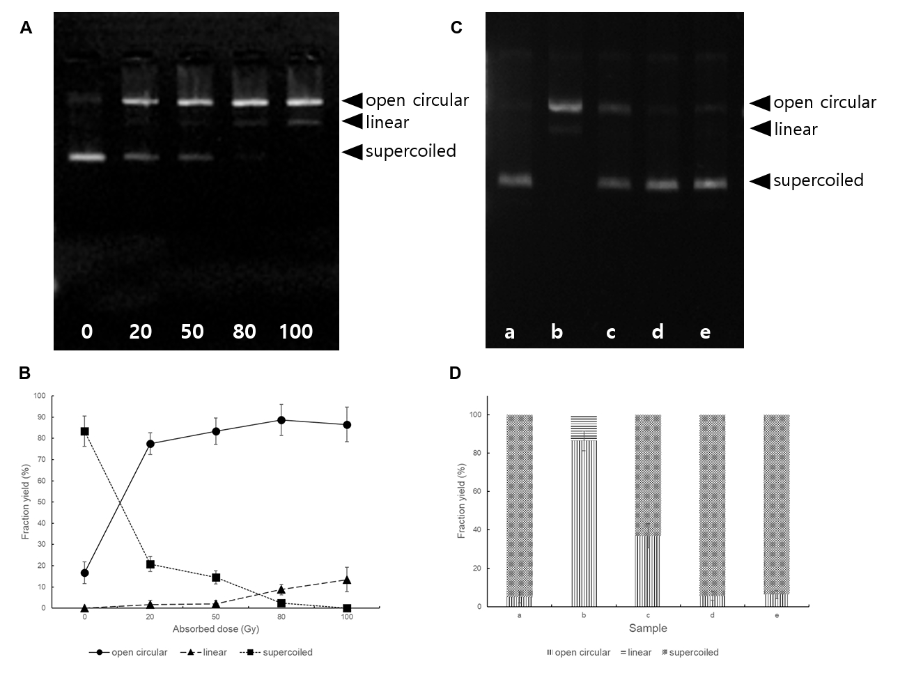
The protective effects of lycopene and β-carotene against gamma-radiation-induced DNA strand breakage were evaluated using purified pUC19 plasmid DNA as an in vitro model. Agarose gel electrophoresis resolved the plasmid into its characteristic conformational forms: the covalently closed supercoiled form, the relaxed open-circular form, and the linear form. The supercoiled form represents predominantly intact plasmid DNA, whereas conversion to the open-circular and linear forms indicates the induction of single-strand breaks and double-strand breaks, respectively (Gual et al., 2011). In this study, non-irradiated pUC19 DNA was detected predominantly in the supercoiled form, with only a small proportion of open-circular DNA, indicating that the purified plasmid was largely intact before irradiation (Fig. 1A). Exposure of pUC19 DNA to gamma radiation produced a clear dose-dependent alteration in plasmid topology. Following irradiation at 20 Gy, the intensity of the supercoiled DNA band decreased, accompanied by a pronounced increase in the open-circular form. This conversion indicates that gamma irradiation at 20 Gy primarily induced single-strand breaks in the plasmid DNA. When the irradiation dose was increased to 80 Gy, a distinct linear DNA band appeared in addition to the open-circular form, while the proportion of the intact supercoiled form was further reduced. The appearance of linear DNA at 80 Gy demonstrates that the accumulation of radiation-induced lesions resulted in breaks affecting both strands of the DNA molecule. Thus, the electrophoretic profile showed a progressive transition from supercoiled DNA to open-circular DNA and subsequently to linear DNA as the absorbed dose increased. The fraction yields calculated from band intensities were shown in Fig. 1B.
The addition of either lycopene or β-carotene before irradiation reduced these changes in plasmid conformation (Fig. 1C). At a concentration of 10 μg/ml (sample c), lycopene partially preserved the supercoiled form and decreased the formation of open-circular relative to the corresponding irradiated sample (sample b) at 100 Gy without carotenoids. A stronger protective effect was observed when the carotenoid concentration was increased to 100 μg/ml. In the presence of 100 μg/ml lycopene or β-carotene (samples d and e, respectively), the electrophoretic pattern of irradiated pUC19 DNA closely resembled that of the non-irradiated control (sample a). The supercoiled form remained the predominant plasmid conformation, whereas the formation of open-circular and linear DNA was markedly suppressed. Collectively, these results demonstrate that lycopene and β-carotene protected plasmid DNA against gamma-radiation-induced strand breakage in a concentration-dependent manner, with 100 μg/ml providing nearly complete protection under the experimental conditions.
The protective effect observed in purified DNA solution is important because this system contains no DNA repair enzymes, transcriptional responses, or cellular antioxidant networks. Therefore, the preservation of plasmid conformation is most consistent with a direct chemical radioprotective action of carotenoids. In aqueous solution, gamma irradiation induces water radiolysis and generates highly reactive species, especially hydroxyl radicals, that attack DNA bases and the sugar-phosphate backbone. Scavenging these radicals before they interact with DNA can reduce the conversion of supercoiled plasmids into open-circular and linear forms. This interpretation is supported by previous studies showing that non-provitamin A carotenoids, including lycopene, are frequently associated with reduced oxidative DNA damage (Azqueta and Collins, 2012).
A classic measured value for E. coli cell volume is 1.1 µm3, with a reported range of 0.44–1.79 µm3 from Coulter-counter/pellet-volume measurements (Kubitschek and Friske, 1986). For mass, a standard E. coli example in exponential growth on glucose minimal medium gives total wet mass = 0.95 pg, calculated from dry mass = 0.28 pg plus water mass = 0.67 pg, assuming ~70% cellular water (Kubitschek, 1986). From these reported values, the concentration of carotenoid in a cell is about 112 µg/ml, when cell accumulates carotenoid at the content of 0.4 mg carotenoid/g dry cell weight. This calculation provides only an approximate equivalence because intracellular carotenoids are not necessarily homogeneously distributed, but can provide rationale for the concentration of carotenoid in DNA solution in this study.
Along with agarose gel electrophoresis, the AFM observations were presented as independent structural confirmation.
AFM analysis of irradiated plasmid DNA
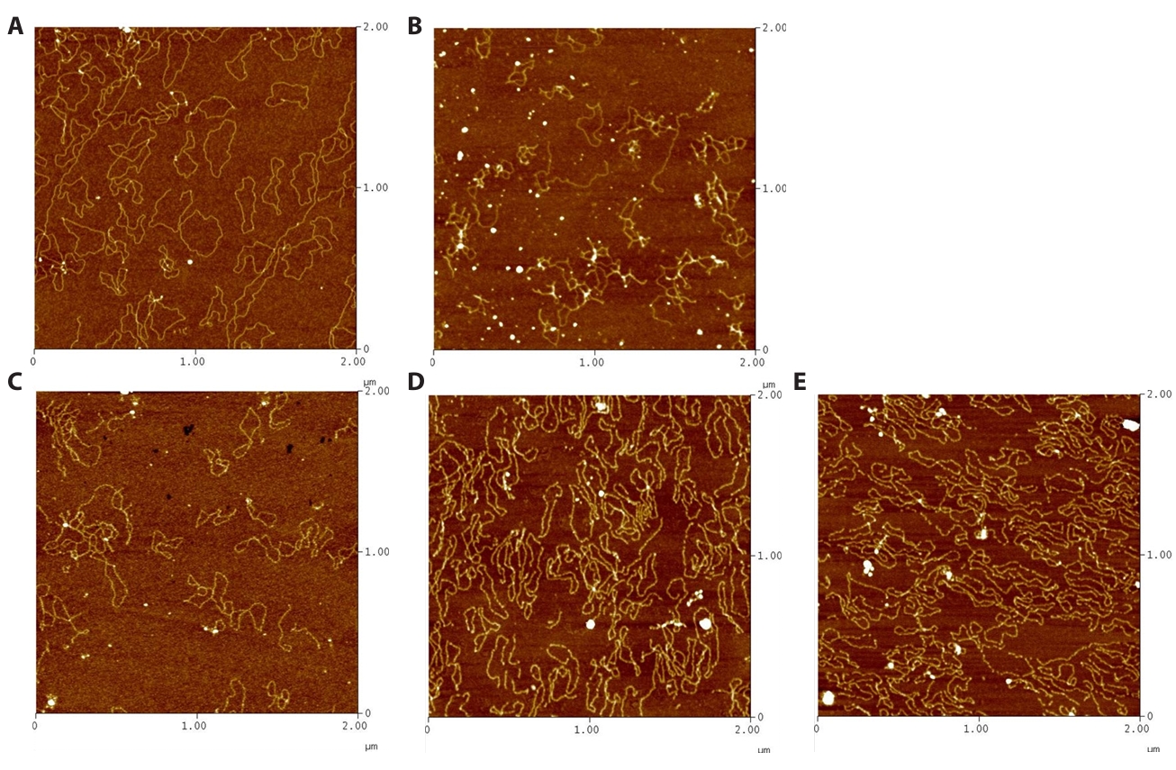
Atomic force microscopy was used to directly examine the structural integrity of the pUC19 plasmid DNA and to confirm the protective effects of lycopene and β-carotene observed by agarose gel electrophoresis (Fig. 2). AFM imaging enabled individual DNA molecules to be visualized at the nanometer scale and provided complementary morphological information regarding radiation-induced changes in plasmid structure. The non-irradiated control sample showed predominantly intact DNA molecules with continuous coiled or circular conformations (Fig. 2A). The DNA contours were clearly defined, and extensive fragmentation was not observed, indicating that the plasmid DNA remained structurally intact during sample preparation and AFM deposition.
In contrast, marked structural alterations were observed in plasmid DNA irradiated in the absence of carotenoids at 100 Gy (Fig. 2B). The irradiated samples contained discontinuous, shortened, and irregular DNA structures rather than the intact circular molecules observed in the non-irradiated control. DNA fragments of different contour lengths were distributed across the substrate, together with partially opened or disrupted plasmid structures. These morphological changes provide direct evidence that gamma irradiation induced substantial damage to the plasmid DNA. The loss of intact circular contours and the appearance of fragmented molecules were consistent with the conversion of supercoiled plasmid DNA into open-circular and linear forms detected by agarose gel electrophoresis. In the electrophoretic analysis, the open-circular form was associated with single-strand breakage, whereas the generation of linear DNA at the higher radiation dose indicated the occurrence of double-strand breaks. AFM therefore provided direct visual support for the dose-dependent disruption of the plasmid structure demonstrated by electrophoretic separation.
The morphology of the irradiated DNA was substantially preserved when lycopene or β-carotene was present during gamma irradiation (Fig. 2C–2E). In carotenoid-treated samples, most DNA molecules retained continuous and intact contours, and circular or coiled plasmid structures remained readily distinguishable. The frequency of visibly fragmented and severely disrupted DNA molecules was markedly lower than that observed in irradiated samples without carotenoids. Although some molecules showed relaxation or minor conformational variation, extensive fragmentation was not apparent. These observations indicate that both lycopene and β-carotene reduced gamma-radiation-induced disruption of the plasmid DNA structure.
The AFM findings were consistent with the concentration-dependent protection detected by agarose gel electrophoresis. At the lower carotenoid concentration of 10 μg/ml, irradiation-induced strand breakage was reduced but was not completely prevented, as indicated by the persistence of a proportion of altered DNA molecules (Fig. 2C). At the carotenoid concentration of 100 µg/ml, the DNA structures observed by AFM were predominantly intact and closely resembled those of the non-irradiated control (Fig. 2D and 2E). Together, these results demonstrate that the protective effects of lycopene and β-carotene were concentration dependent and were detectable using two methodologically distinct approaches.
AFM has previously been applied to demonstrate that DNA-damaging oxidative conditions caused concentration-dependent alterations in plasmid conformation and that severe damage resulted in the loss of circular structures and the appearance of linear fragments (Dai et al., 2022). The same study showed that the presence of a protective antioxidant-compatible solute markedly reduced DNA fragmentation, illustrating the usefulness of AFM for directly evaluating molecular protection against oxidative DNA damage. Although the damaging agent and protective compound investigated by previous report differed from those used in the present study, the reported morphological progression from intact circular DNA to disrupted and fragmented molecules support the interpretation of the present AFM observations.
Discussion
Gamma irradiation damages biological molecules through direct and indirect mechanisms. Direct effects occur when radiation energy is deposited directly in DNA or other macromolecules, whereas indirect effects result from reactive species generated by water radiolysis.
In hydrated cells, ionizing radiation damages DNA predominantly through reactive oxygen species (ROS) generated by water radiolysis (Azzam et al., 2012). Unrepaired lesions, such as strand breaks and stalled replication forks, generate single-stranded DNA (ssDNA) that nucleates RecA filament formation, promotes LexA autocleavage, and induces the SOS response (Little and Mount, 1982). RecA-mediated homologous recombination, supported by the RecFOR pathway, is central to rejoining gamma-induced double-strand breaks, whereas persistent lesions may recruit error-prone translesion polymerases and elevate mutagenesis (Goodman and Woodgate, 2013). Because carotenoids efficiently quench singlet oxygen and scavenge peroxyl radicals (Stahl and Sies, 2003), the enhanced survival of carotenoid-accumulating E. coli is most plausibly explained not by the absence of DNA damage but by a shift in the balance between damage and repair: carotenoids act upstream to lower the ROS-derived lesion burden, thereby reducing the load on the cellular repair network. Carotenoid accumulation therefore represents an additional, prevention-oriented layer of protection that complements the intrinsic DNA-repair capacity of the cell.
The present findings support this mechanism at both cellular and molecular levels. In recombinant E. coli, carotenoid accumulation increased D10 values several-fold, indicating improved survival after gamma irradiation.
Because gamma rays are highly penetrating, membrane-associated carotenoids cannot act as physical shields, and the extremely short diffusion radius of hydroxyl radicals means that membrane-localized carotenoids are unlikely to scavenge •OH generated directly at the nucleoid. Radioprotection by carotenoids in cells is therefore better attributed to suppression of secondary, more diffusible reactive species and radical chain reactions—most notably chain-breaking inhibition of membrane lipid peroxidation and quenching of singlet oxygen and peroxyl radicals—which lowers the global intracellular oxidative burden rather than protecting DNA through direct co-localized scavenging.
In purified plasmid DNA, lycopene and β-carotene reduced the conversion of supercoiled DNA to open-circular and linear forms and preserved intact DNA morphology in AFM images. Because the purified DNA assay lacks cellular repair systems, the observed protection is most likely due to direct scavenging of radiation-derived radicals or interruption of radical chain reactions near the DNA molecule.
Accordingly, the cell-free assay should be interpreted as demonstrating the intrinsic radical-scavenging capacity of carotenoids toward DNA, whereas the enhanced survival of recombinant cells is most likely mediated by prevention of lipid peroxidation and a reduction in the diffusible intracellular oxidative burden, rather than by direct shielding of the nucleoid.
AFM was employed for qualitative, single-molecule visualization of plasmid integrity, consistent with prior AFM studies of DNA damage (Dai et al., 2022). Quantitative fractions of the supercoiled, open-circular, and linear forms were determined by densitometric analysis of agarose-gel bands (Fig. 1B and 1D), which provided the quantitative complement to the AFM observations.
The use of recombinant E. coli is a key strength of this study. Previous work with naturally radioresistant bacteria, particularly D. radiodurans, has suggested that carotenoids contribute to resistance, but the interpretation is complicated by highly efficient DNA repair systems, proteome protection, antioxidant enzymes, and other specialized adaptations (Ji, 2010; Slade and Radman, 2011; Sun et al., 2009; Tian et al., 2007). By introducing carotenoid biosynthesis into a radiation-sensitive host, the present study provides a more controlled system for evaluating the contribution of carotenoid accumulation. The observation that plasmid-bearing strains did not show increased resistance under carotenoid-producing conditions further indicates that the protective effect was most likely attributable primarily to carotenoid accumulation rather than to the recombinant plasmid background alone. Because heterologous pathway expression and metabolic load may exert additional physiological effects, carotenoid accumulation is best regarded as the most likely major contributor to the observed protection rather than the sole factor.
We emphasize that these results do not imply that carotenoids act independently of DNA repair. Rather, carotenoid accumulation appears to reduce the initial oxidative lesion burden, thereby lessening the load on the cellular repair systems with which it acts in concert. Definitive genetic dissection of the relative contributions of prevention and repair will require repair-deficient (e.g., recA) backgrounds, which we identify as an important direction for future study.
Overall, these results suggest that carotenoid biosynthesis can be used as a metabolic strategy to enhance microbial tolerance to ionizing radiation. This finding has potential significance for microbial biotechnology, food and environmental microbiology, and the development of stress-tolerant production strains. It also provides a useful experimental framework for comparing the radioprotective activities of different carotenoid structures in a common host background.
Acknowledgments
The research was supported by the Chonnam National University Supporting Program (2026-1416-01) and the KAERI institutional R&D Program (Project No. 523510-26) funded by the Ministry of Science and ICT (MIST).
Conflict of Interest
The authors declare that they have no competing interests.
Ethical Statements
Not applicable
Supplementary Information
The online version contains supplementary material available at https://doi.org/10.71150/jm.2606016
Fig. S1.
Survival curves of recombinant E. coli strains with/without carotenoid accumulation. (A) E. coli DH5α, (B) E. coli DH5α(pTrc99A), (C) E. coli DH5α(pT-LYCm4), and (D) E. coli DH5α(pT-HB) in LB medium with/without Ap. And, (E) E. coli DH5α, (F) E. coli DH5α(pTrc99A), (G) E. coli DH5α(pT-LYCm4), and (H) E. coli DH5α(pT-HB) in 2YT + 0.5% glycerol with/without Ap.
jm-2606016-Supplementary-Fig-S1.pdf
Fig. 1.(A) The image of agarose gel electrophoresis and (B) plot of the fraction yields of different forms for pUC19 DNA irradiated at different doses of 20, 50, 80, and 100 Gy, respectively. (C) The image of agarose gel electrophoresis and (D) plot of the fraction yields of different forms for pUC19 DNA treated with (a) non-irradiation, (b) irradiation at 100 Gy, (c) irradiation at 100 Gy with 10 μg lycopene/ml, (d) irradiation with 100 Gy with 100 μg lycopene/ml, and (e) irradiation with 100 Gy with 100 μg β-carotene/ml, respectively. Error bars represent standard deviation.
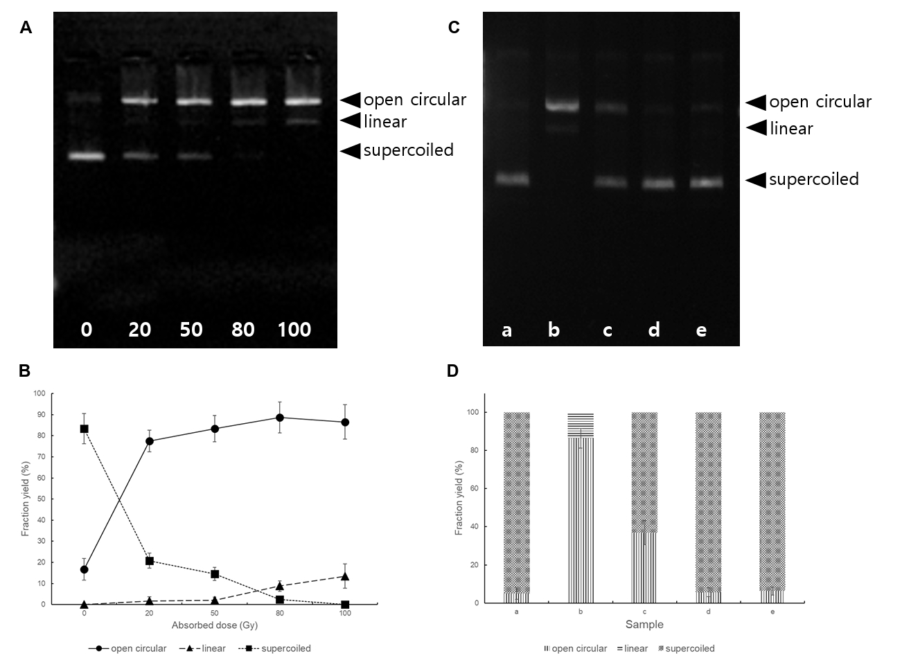
Fig. 2.AFM images of plasmid pUC19 DNA in deposition buffer (1 mM MgCl2, 10 mM HEPES; pH 7.5). (A) pUC19 DNA before irradiation, (B) pUC19 DNA irradiated with 100 Gy without carotenoid, (C) pUC19 DNA irradiated with 100 Gy with 10 μg lycopene/ml, (D) pUC19 DNA irradiated with 100 Gy with 100 μg lycopene/ml, and (E) pUC19 DNA irradiated with 100 Gy with 100 μg β-carotene/ml, respectively.
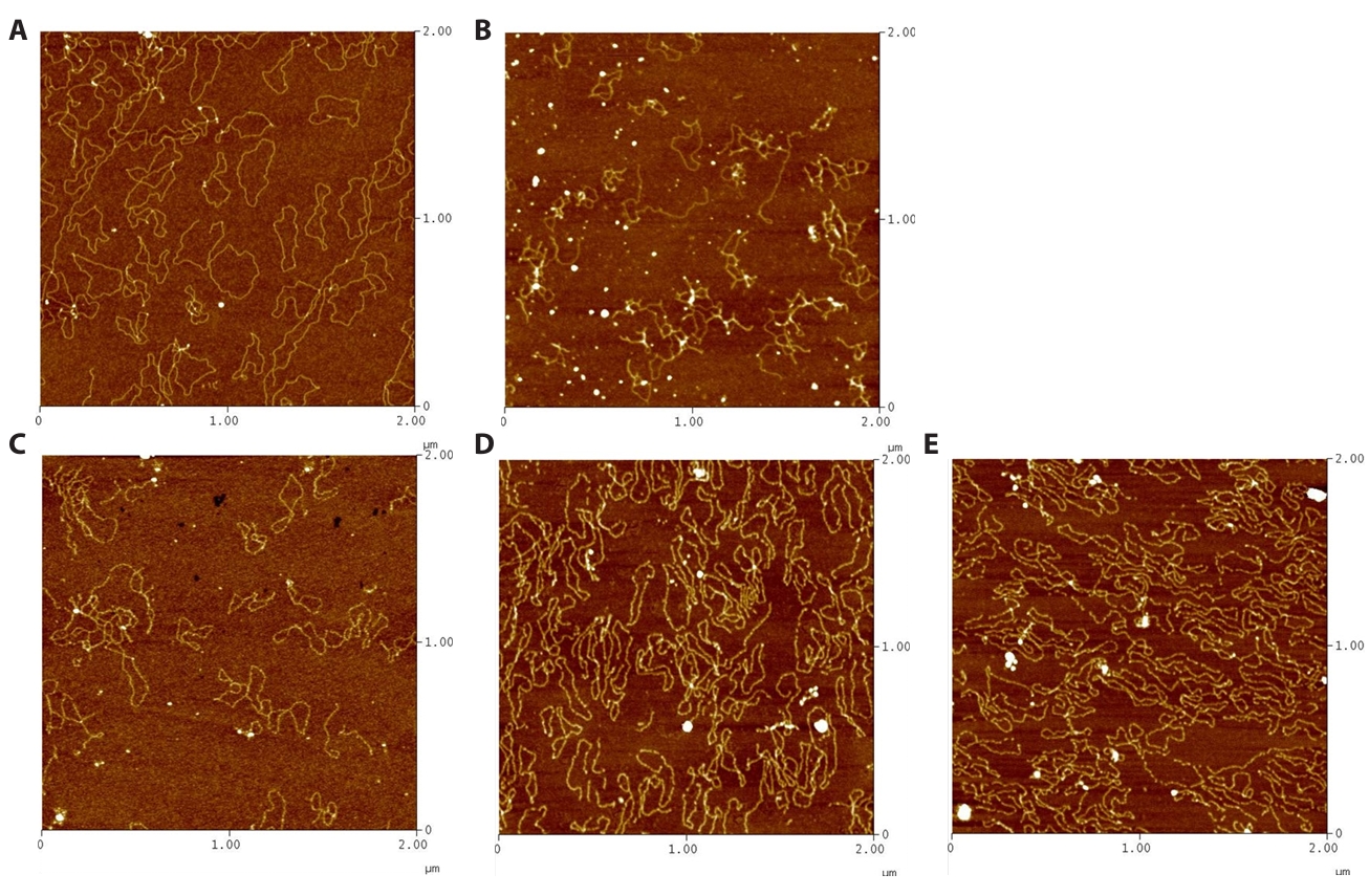
Table 1.The lists of recombinant E. coli strains and plasmids used in this study
|
Bacterial strains and plasmids |
Characteristics |
References |
|
E. coli DH5α |
|
|
|
E. coli DH5α (pT-LYCm4) |
Lycopene producer |
Yoon et al. (2006)
|
|
E. coli DH5α (pT-HB) |
β-Carotene producer |
Yoon et al. (2007)
|
|
pTrc99A |
4,176 bp |
Pharamacia |
|
pUC19 |
2,686 bp |
New England Biolabs |
Table 2.D10 values of various recombinant E. coli strains with/without carotenoid accumulation
|
Strain |
D10 value (Gy) |
Culture condition |
Carotenoid content (mg/g cell weight) |
|
E. coli DH5α |
91.4 ± 11.9a
|
LB |
- |
|
E. coli DH5α (pTrc99A) |
97.57 ± 15.2a
|
LB + Ap |
- |
|
E. coli DH5α (pT-LYCm4) |
106.4 ± 14.6a
|
LB + Ap |
- |
|
E. coli DH5α (pT-HB) |
98.2 ± 15.7a
|
LB + Ap |
- |
|
E. coli DH5α |
99.8 ± 16.1a
|
2YT + 0.5% glycerol |
- |
|
E. coli DH5α (pTrc99A) |
112.25 ± 21.1a
|
2YT + 0.5% glycerol + Ap |
- |
|
E. coli DH5α (pT-LYCm4) |
295.4 ± 49.1b
|
2YT + 0.5% glycerol + Ap |
0.52 ± 0.26 |
|
E. coli DH5α (pT-HB) |
358.4 ± 51.2b
|
2YT + 0.5% glycerol + Ap |
0.43 ± 0.13 |
References
- Ahn JW, Choi JI. 2026. Recent advances in Chlamydomonas reinhardtii as a green factory for sustainable biofuels and bioproduct production. N Biotechnol. 93: 362–377. Article
- Azqueta A, Collins AR. 2012. Carotenoids and DNA damage. Mutat Res. 733: 4–13. ArticlePubMed
- Azzam EI, Jay-Gerin JP, Pain D. 2012. Ionizing radiation-induced metabolic oxidative stress and prolonged cell injury. Cancer Lett. 327: 48–60. ArticlePubMed
- Battista J. 1997. Against all odds: The survival strategies of Deinococcus radiodurans. Annu Rev Microbiol. 51: 203–224. ArticlePubMed
- Choi JI, Lee HS, Kim JH, Lee KW, Lee JW, et al. 2008. Controlling the radiation degradation of carboxymethylcellulose solution. Polym Degrad Stab. 93: 310–315. Article
- Dai T, Wang Y, Yang G. 2022. Visualization of DNA damage and protection by atomic force microscopy in liquid. Int J Mol Sci. 23: 4388.ArticlePubMedPMC
- D'Auria Vieira de Godoy PR, Nakamura A, Khavari AP, Sangsuwan T, Haghdoost S. 2021. Effect of dose and dose rate of gamma irradiation on the formation of micronuclei in bone marrow cells isolated from whole-body-irradiated mice. Environ Mol Mutagen. 62: 422–427. ArticlePubMedLink
- Farci D, Slavov C, Tramontano E, Piano D. 2016. The S-layer protein DR_2577 binds deinoxanthin and under desiccation conditions protects against UV-radiation in Deinococcus radiodurans. Front Microbiol. 7: 155.ArticlePubMedPMC
- Goodman MF, Woodgate R. 2013. Translesion DNA polymerases. Cold Spring Harb Perspect Biol. 5: a010363.ArticlePubMedPMC
- Gual MR, Milian FM, Deppman A, Coelho PR. 2011. Study of DNA damage with a new system for irradiation of samples in a nuclear reactor. Appl Radiat Isot. 69: 373–376. ArticlePubMed
- Ji HF. 2010. Insight into the strong antioxidant activity of deinoxanthin, a unique carotenoid in Deinococcus radiodurans. Int J Mol Sci. 11: 4506–4510. ArticlePubMedPMC
- Krisko A, Radman M. 2010. Protein damage and death by radiation in Escherichia coli and Deinococcus radiodurans. Proc Natl Acad Sci USA. 107: 14373–14377. ArticlePubMedPMC
- Kubitschek HE. 1986. Increase in cell mass during the division cycle of Escherichia coli B/rA. J Bacteriol. 168: 613–618. ArticlePubMedPMCLink
- Kubitschek H, Friske J. 1986. Determination of bacterial cell volume with the Coulter Counter. J Bacteriol. 168: 1466–1467. ArticlePubMedPMCLink
- Lim S, Jung JH, Blanchard L, de Groot A. 2019. Conservation and diversity of radiation and oxidative stress resistance mechanisms in Deinococcus species. FEMS Microbiol Rev. 43: 19–52. ArticlePubMedPMC
- Little JW, Mount DW. 1982. The SOS regulatory system of Escherichia coli. Cell. 29: 11–22. ArticlePubMed
- Makarova KS, Aravind L, Wolf YI, Tatusov RL, Minton KW, et al. 2001. Genome of the extremely radiation-resistant bacterium Deinococcus radiodurans viewed from the perspective of comparative genomics. Microbiol Mol Biol Rev. 65: 44–79. ArticlePubMedPMCLink
- Maoka T. 2020. Carotenoids as natural functional pigments. J Nat Med. 74: 1–16. ArticlePubMedPDF
- Moreira RG, Puerta-Gomez AF, Kim J, Castell-Perez ME. 2012. Factors affecting radiation D-values (D10) of an Escherichia coli cocktail and Salmonella Typhimurium LT2 inoculated in fresh produce. J Food Sci. 77: E104–E111. ArticlePubMed
- Park HS, Choi JI. 2026. Characterization and transcriptomic analysis of Antarctic Planococcus sp. mutant with enhanced carotenoid content. J Microbiol Biotechnol. 36: e2512033. ArticlePubMedPMC
- Park SH, Sohn YJ, Park SJ, Choi JI. 2020. Effect of DR1558, a Deinococcus radiodurans response regulator, on the production of GABA in the recombinant Escherichia coli under low pH conditions. Microb Cell Fact. 19: 64.ArticlePubMedPMCPDF
- Reis-Mansur M, Cardoso-Rurr JS, Silva J, de Souza GR, Cardoso VDS, et al. 2019. Carotenoids from UV-resistant Antarctic Microbacterium sp. LEMMJ01. Sci Rep. 9: 9554.ArticlePubMedPMCPDF
- Rueden CT, Schindelin J, Hiner MC, DeZonia BE, Walter AE, et al. 2017. ImageJ2: ImageJ for the next generation of scientific image data. BMC Bioinformatics. 18: 529.ArticlePubMedPMCPDF
- Sandmann G. 2019. Antioxidant protection from UV- and light-stress related to carotenoid structures. Antioxidants (Basel). 8: 219.ArticlePubMedPMC
- Slade D, Radman M. 2011. Oxidative stress resistance in Deinococcus radiodurans. Microbiol Mol Biol Rev. 75: 133–191. ArticlePubMedPMCLink
- Stahl W, Sies H. 2003. Antioxidant activity of carotenoids. Mol Aspects Med. 24: 345–351. ArticlePubMed
- Sun Z, Shen S, Tian B, Wang H, Xu Z, et al. 2009. Functional analysis of γ-carotene ketolase involved in the carotenoid biosynthesis of Deinococcus radiodurans. FEMS Microbiol Lett. 301: 21–27. ArticlePubMed
- Tian B, Hua Y. 2010. Carotenoid biosynthesis in extremophilic Deinococcus-Thermus bacteria. Trends Microbiol. 18: 512–520. ArticlePubMed
- Tian B, Xu Z, Sun Z, Lin J, Hua Y. 2007. Evaluation of the antioxidant effects of carotenoids from Deinococcus radiodurans through targeted mutagenesis, chemiluminescence, and DNA damage analyses. Biochim Biophys Acta. 1770: 902–911. ArticlePubMed
- Yoon SH, Lee YM, Kim JE, Lee SH, Lee JH, et al. 2006. Enhanced lycopene production in Escherichia coli engineered to synthesize isopentenyl diphosphate and dimethylallyl diphosphate from mevalonate. Biotechnol Bioeng. 94: 1025–1032. ArticlePubMed
- Yoon SH, Park HM, Kim JE, Lee SH, Choi MS, et al. 2007. Increased β-carotene production in recombinant Escherichia coli harboring an engineered isoprenoid precursor pathway with mevalonate addition. Biotechnol Prog. 23: 599–605. ArticlePubMed
Citations
Citations to this article as recorded by

 MSK




 ePub Link
 Cite this Article
 XML Download