ABSTRACT
- Plastic waste in marine environments provides novel habitats for diverse organisms, forming distinct microbial ecosystems known as the ‘plastisphere’. Although bacteria of the plastisphere have been widely studied, the role of fungi in plastisphere formation and plastic degradation remains largely unexplored. Thus, we investigated temporal changes in culturable fungal community composition on three common plastic types—high-density polyethylene, low-density polyethylene, and polypropylene—across the early (seven days) and mature (30 days) plastisphere developmental stages through a marine mesocosm experiment. In total, 436 fungal strains were isolated and identified as belonging to 179 taxa, with Penicillium, Cladosporium, Trichoderma, Aspergillus, and Fusarium as the dominant genera. Temporal shifts in the species richness of the dominant genera were observed: Cladosporium showed higher species richness at the early stage, whereas that of Trichoderma increased at the mature stage. Plastic degradation assays revealed that 54.6% of the strains exhibited measurable degradation capacity, with patterns varying by plastic type rather than fungal developmental stage. Scanning electron microscope observations revealed surface damage patterns including cracks, pitting, and erosion on the plastic surfaces. These findings provide novel insights into the composition and functional heterogeneity of culturable fungal communities in the marine plastisphere and suggest that plastisphere fungi play diverse ecological roles beyond direct plastic degradation.
-
Keywords: culturable fungi, marine plastisphere, fungal community, plastic degradation, mesocosm, biofilm succession
Introduction
Plastic waste accumulation extensively impacts marine ecosystems (Yu et al., 2023), as 75–199 million tons of plastic waste are estimated to flow into the marine environment (UNEP, 2021). High-density polyethylene (HDPE), low-density polyethylene (LDPE), and polypropylene (PP) account for the highest global plastic production volumes (Geyer et al., 2017). Plastic wastes provide a novel habitat for a variety of organisms, which is referred to as the ‘plastisphere.’ This habitat is distinct from those of the surrounding marine microbial communities and provides a unique ecological niche through interactions with the surrounding seawater conditions (Amaral-Zettler et al., 2020; Jacquin et al., 2019; Li et al., 2024; Zettler et al., 2013).
When plastics are exposed to the marine environment, a conditioning film rapidly forms on their surfaces (Rummel et al., 2021), enabling pioneer microbes, such as bacteria and diatoms, to develop an extracellular polymeric substance (EPS)-based plastisphere, followed by the adhesion of secondary colonizers, such as ciliates, dinoflagellates, and fungi (Dobretsov et al., 2013; Qian et al., 2022). As succession progresses, sessile marine organisms such as macroalgae, barnacles, and tube worms also settle, leading to the maturation of the plastisphere (Dobretsov et al., 2013). Importantly, early microbial communities do not only initiate plastisphere formation through EPS production but may also modulate plastic surface properties and resource availability, thereby influencing both degradation processes and colonization toward the development of a mature plastisphere. Although bacterial succession on marine plastics has been studied alongside biofilm development and functional activity profiles, including increases in extracellular enzymatic activity during prolonged immersion (Odobel et al., 2021), the roles and dynamics of other microbial groups are yet to be systematically characterized.
Fungi are efficient decomposers of recalcitrant substrates (e.g., lignin and cellulose), suggesting their potential for degrading marine plastic waste. Previous research on fungi associated with the marine plastisphere has primarily focused on identifying a few strains with high plastic degradation capacities (Berger et al., 2026; Vaksmaa et al., 2024) or characterizing their plastic degradation mechanisms (Gao et al., 2022; Zhang et al., 2022). However, studies that simultaneously track temporal shifts in the compositions of marine plastisphere fungal communities and the corresponding changes in plastic degradation capacity remain limited.
Plastisphere succession has been reported to enrich degradation-related taxa and functions over time (Wang et al., 2022), and plastic-utilizing fungal isolates have more frequently been recovered after longer immersion periods (Philippe et al., 2024). Based on these observations, we hypothesized that fungal strains with strong degradation capabilities would appear more frequently during the mature stage of plastisphere development. We aimed to investigate the temporal changes in fungal community composition in marine plastispheres by comparing the culturable fungal diversity on plastic sheets submerged for seven days (early stage) and 30 days (mature stage) using a mesocosm experiment. The plastic degradation capacity of each fungal strain was evaluated through weight-loss measurements and scanning electron microscope (SEM) observations to directly assess plastic surface damage. By integrating these results, this study provides insights—including the discovery of fungal resources with the potential for bioremediation—into fungal contributions to the formation and maintenance of the marine plastisphere.
Materials and Methods
Mesocosm design and sample collection
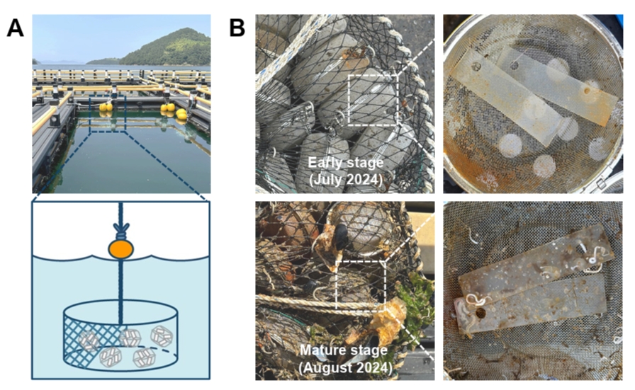
Mesocosm experiments were conducted from July to August 2024 in a sea cage at the South Sea Research Institute of the Korea Institute of Ocean Science and Technology (KIOST) (34°59’33.5” N, 128°40’27.0” E). The detailed layout of the mesocosm structure is shown in Fig. 1A. Briefly, each mesocosm structure consisted of stainless-steel tea balls (prepared for each plastic type) containing four plastic sheet pieces internally connected by sterilized fishing lines, which were connected to cylindrical pots. To minimize wave disturbance, each mesocosm structure was submerged at approximately 1 m below the sea surface using buoys.
For the preparation of the mesocosm structures, three types of plastic sheets of 1 mm thickness were purchased: HDPE (Goodfellow, catalogue no. ET32-SH-000100), LDPE (ET31-SH-000110), and PP (PP30-SH-000100). Plastic sheets were cut into 8 × 2 cm pieces and surface-sterilized by sequential washing with 70% (v/v) ethanol (Samchun Chemicals, Republic of Korea) and distilled water, followed by overnight exposure to UV light (253.7 nm). Stainless-steel tea balls and fishing lines were sterilized by autoclaving.
One unit of each mesocosm was collected at seven and 30 days after deployment (one unit per plastisphere developmental stage). The plastic sheets in each tea ball were rinsed three times on site with sterilized distilled water (DW) and subjected to different pretreatments, depending on the subsequent experimental objectives. For the fungal isolation experiments, each plastic sheet (nine replicates/plastic type) was immersed in a modified fungal inoculation solution to maintain fungal cell viability and facilitate cell detachment. The solution consisted of 30% (v/v) glycerol (Junsei Chemical, Japan) and 0.05% (w/v) agar (BD DifcoTM, Detroit, USA) prepared in sterilized seawater (SSW; instead of distilled water) supplemented with 0.05% (v/v) Tween 80 (Sigma-Aldrich, USA) as a surfactant. For the microscopic observation of plastisphere colonization and plastic surface alterations, each plastic sheet (two replicates/plastic type) was immersed in SSW. All pretreated samples were transported to the laboratory at 4℃.
Fungal isolation
The sampled plastic sheets were immersed in the modified fungal inoculation solutions and vortexed at maximum speed for 10 min to detach biofilms from the plastic sheet surfaces. For fungal isolation, the resulting homogenates were filtered through 0.2-μm polycarbonate track-etched membrane filters (GVS Filter Technology, USA) to collect fungal cells. The membrane filters were then placed onto three different isolation media: marine agar (MA; BD DifcoTM, USA), minimal medium (MM; 2.0 g NaNO3, 0.7 g KH2PO4, 0.3 g K2HPO4, 0.5 g KCl, 0.5 g MgSO4·7H2O, 0.1 g FeSO4·7H2O, and 30 g agar/L of SSW) (Mathur and Prasad, 2012), and yeast extract peptone dextrose agar (YPDA; BD DifcoTM, USA). For each plastic type, three replicate filters were used, with one filter placed on each medium type and incubated at 25℃ for two weeks. To obtain pure cultures, single colonies were subcultured on fresh potato dextrose agar (PDA; BD DifcoTM, USA) supplemented with SSW. Each pure fungal culture was preserved as 20% glycerol stocks supplemented with SSW and stored at –20℃ in the Seoul National University Fungal Collection.
Molecular identification
Genomic DNA was extracted from the fresh mycelia of each PDA fungal culture using an AccuPrep Genomic DNA Extraction kit (Bioneer, Republic of Korea), according to the manufacturer’s instructions. Polymerase chain reaction (PCR) amplifications were performed in a C1000 Thermal Cycler (Bio-Rad Laboratories, USA) using AccuPower® PCR Premix (Bioneer, Republic of Korea). The internal transcribed spacer (ITS) region sequences were amplified using fungal-specific primers (PCR conditions are summarized in Table S1). PCR amplicons were verified by electrophoresis on 1% agarose gel and purified using an ExoSAP-IT Express PCR Product Cleanup kit (Thermo Fisher Scientific, USA). Sequencing was performed by Macrogen (Republic of Korea) on an ABI PRISM 3700 Genetic Analyzer (Life Technologies, USA) using the same primer sets used for PCR.
The sequences obtained were proofread, trimmed, and manually edited using Geneious Prime 2024.7.12 (https://www.geneious.com). Forward and reverse reads were assembled using the de novo assembly function in Geneious Prime to obtain consensus sequences. Preliminary identification of higher taxonomic ranks (genus level or above) was performed using NCBI BLAST searches of the ITS sequences. For taxa that were not reliably identifiable to the species level based on the ITS sequence alone, additional genetic markers were amplified and sequenced under the conditions specified in Table S1. The newly generated sequences were deposited in the GenBank database of the NCBI (accession numbers are listed in Table S2).
Reference sequences were retrieved from GenBank based on a literature search and combined with our newly obtained sequences for phylogenetic analysis. A phylogenetic tree for each genus was inferred using either (1) FunVIP v.0.4.1 with “accurate” mode and 1,000 bootstrap replicates (Seo et al., 2025), or (2) RAxML implemented in Geneious Prime with 1,000 bootstrap replicates. The final species-level identification of each strain was based on the resulting phylogeny.
Assessment of plastic degradation capacity by weight-loss measurements
The plastic-degrading capacity of each fungal isolate was evaluated individually using an in vitro degradation assay. To assess the plastic degradation capacities of fungi, 6 mm diameter fungal mycelial discs cut from one-week-old PDA fungal cultures supplemented with SSW were inoculated at the center of MM plates. Plastic sheets (HDPE, LDPE, and PP) were punched into discs of 6 mm in diameter and surface-sterilized using the same methods as those used for the mesocosm plastic sheets. Sterilized plastic discs were placed approximately 15 mm from each fungal inoculum and served as the sole carbon source. Each strain was tested only against the plastic type from which it was originally isolated. After 60 days of incubation at 25℃, plastic discs were washed to remove adherent mycelia by sequential washing with 30% (w/v) sodium dodecyl sulfate (Sigma-Aldrich, USA), 70% (v/v) ethanol (Samchun Chemicals, Republic of Korea), and DW (Sathiyabama et al., 2024). Subsequently, the discs were oven-dried at 40°C for two to three days, and each disc was weighed using an electronic balance (EPG213C, Ohaus Co., USA). The control samples consisted of plastic discs without fungal inoculation that underwent identical incubation, washing, and measurement procedures.
Plastic degradation capacity was assessed by weight-loss percentage, calculated as
with all values presented as Mean ± Standard Deviation relative to the controls. In cases where experimental discs showed weight gain relative to controls, the weight loss percentage (%) was adjusted to 0.
Scanning electron microscopic analysis
To visually examine plastic degradation patterns by fungi, plastic samples were analyzed using a field-emission SEM (FE-SEM) at the National Instrumentation Center for Environmental Management (NICEM), Seoul National University (Republic of Korea). Two types of plastic samples were analyzed: (1) plastic sheets from mesocosm experiments (two sheets/plastic type at each stage) and (2) plastic discs from degradation assays inoculated with the fungal strain showing the highest degradation capacity. For the latter samples (plastic discs), adherent mycelia were removed prior to fixation by sequential washing with 30% (w/v) sodium dodecyl sulfate (SDS; Sigma-Aldrich, USA), 70% ethanol, and DW (with vortexing for 20 min after each step). All samples were subsequently fixed in 3% (v/v) glutaraldehyde (Thermo Fisher Scientific, USA) for 24–48 h and rinsed three times in DW at room temperature (10 min/wash). Fixed samples were dehydrated using a graded ethanol series (30%, 50%, 70%, 80%, and 90%; 10 min in each solution, with two final exchanges at 100%), followed by one treatment with a 1:1 (v/v) mixture of ethanol and hexamethyldisilazane (HMDS; Sigma-Aldrich, USA) and three with 100% (v/v) HMDS (10 min each). The samples were then air-dried for 48 h, sputter-coated with platinum using a sputter coater (EM ACE200, Leica, Austria), and analyzed using FE-SEM (SIGMA, Carl Zeiss, UK) at an accelerating voltage of 5.0 kV.
Statistical analysis
Fungal communities were analyzed using R v.4.5.0. Diversity analyses were conducted with plastic type (HDPE, LDPE, or PP) and plastisphere developmental stage (early or mature) as fixed factors. Fungal community composition was characterized, diversity metrics were calculated using the vegan package v.2.7.1, and plots were generated using ggplot2 v.3.5.2 (Dixon, 2003; Wickham, 2011). For alpha diversity, the inverse Simpson and exponential Shannon indices were calculated and compared using nonparametric tests (Hill, 1973; Hurlbert, 1971; Lande, 1996). Differences in alpha diversity between developmental stages were assessed using the paired Wilcoxon signed-rank test, whereas those among plastic types were assessed using the Friedman test (Friedman, 1937; Wilcoxon, 1945). P-values were adjusted for multiple comparisons using the Benjamini–Hochberg procedure, and the adjusted p-values were reported as q-values (Benjamini and Hochberg, 1995). Beta diversity was assessed using the Jaccard index, and principal coordinates analysis (PCoA) was performed on the resulting distance matrix with the Lingoes correction to account for negative eigenvalues (Gower, 1966). Heat-tree visualizations were performed using the metacoder package v.0.3.8 (Foster et al., 2017).
Results
Visual characterization of plastisphere developmental stages
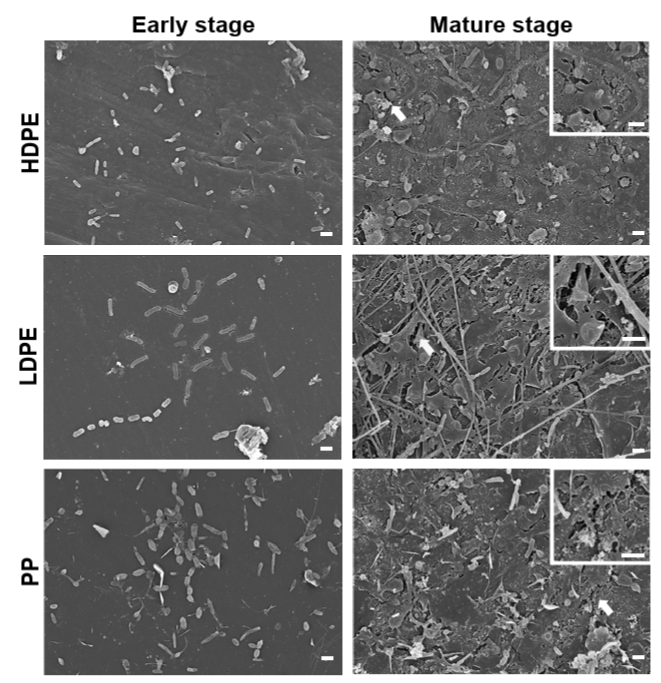
The mesocosm structures sampled at each plastisphere developmental stage showed clear differences in colonization patterns. Macroscopically, the early mesocosm structure appeared relatively clean with minimal surface colonization, whereas the mature mesocosm structure exhibited conspicuous and dense colonization by diverse marine organisms, such as macroalgae (e.g., Ulva, Cladophora, and Pylaiella) and sessile marine fauna, including Pacific oysters, Mediterranean mussels, barnacles, and bryozoans (Fig. 1). Microscopically, the plastic sheet surfaces exhibited increasingly complex patterns over time, consistent with the macroscopic changes. SEM observations revealed that early-stage plastic surfaces were sparsely colonized and only bacterial morphotypes were present. By contrast, mature-stage plastic sheets were thoroughly covered with rod-shaped and filamentous microorganisms, which were intricately intertwined with particulate matter. Notably, signs of surface degradation, including cracks and pitting, were observed across all plastic types in the mature stage (Fig. 2).
Culturable fungal diversity in marine plastisphere communities
In total, 436 fungal strains were isolated from the plastic sheets sampled at the two plastisphere developmental stages: 214 and 222 strains from the early (seven days) and mature (30 days) stages, respectively. By plastic type, 132 strains were isolated from HDPE (early stage, 54; mature stage, 78), 161 from LDPE (early stage, 95; mature stage, 66), and 143 from PP (early stage, 65; mature stage, 78). ITS sequences were successfully obtained from all 436 strains, and BLAST searches identified 179 taxa, of which 178 were assigned to 57 known genera; one taxon (strain number SFC20240812-M088) remained unassigned, only possibly matching with “Microascales sp.” (Accession Number PZ123891). Additional genetic markers were sequenced for species-level identification, upon which 452 sequences were obtained: 23 LSU, 4 SSU, 37 ACT, 41 CAM, 5 GAPDH, 1 RPB1, 17 RPB2, 193 TEF1, and 131 TUB2. By using phylogenetic analyses with multi-locus datasets, 179 taxa were identified as belonging to 57 genera, 37 families, eight classes, and four phyla (data not shown).
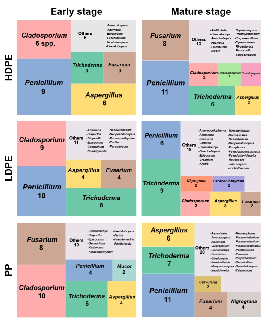
The fungal community composition differed across the developmental stages and plastic types (Fig. 3). In the early stage, a few genera dominated the communities, whereas the diversity increased toward the mature stage. Species richness was higher in the mature stage than in the early stage, and temporal changes in species richness across the three major genera (Penicillium, Cladosporium, and Trichoderma) were consistent across the plastic types. Penicillium consistently exhibited a relatively high species richness in both stages across all plastic types, but particularly so in HDPE. Cladosporium showed higher species richness at the early stage across all plastic types, but decreased with maturation, whereas Trichoderma showed an opposite trend. Aspergillus and Fusarium also showed high species richness, with the relative richness varying across plastic types and developmental stages.
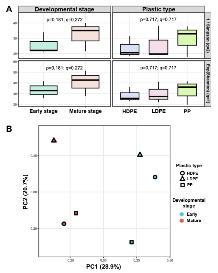
Alpha diversity indices (inverse Simpson and exponential Shannon) were higher in the mature than in the early stage (Fig. 4A). The fungal communities on PP showed higher species richness than those on HDPE and LDPE, although not significantly so. PCoA based on the Jaccard index revealed some separation in the beta diversity between developmental stages along the two main components, where the first and second principal components explained 28.9% and 20.7% of the total variance, respectively (Fig. 4B). However, no clear clustering was observed by plastic type.
Plastic degradation assay results
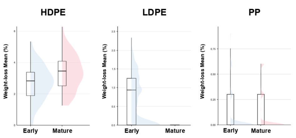
Of the 436 fungal strains isolated, 238 (54.6%) showed measurable plastic degradation capacity compared to the same plastic type from which they were initially isolated. The weight-loss percentages of plastics were measured after 60 days of incubation and those of strains isolated from plastics in the early and mature stages were compared (Fig. 5). Among strains isolated from HDPE, LDPE, and PP, those from the early stage degraded plastic by 0.94–5.35%, 0.31–2.34%, and 0.30–0.90%, respectively; whereas those from the mature stage degraded plastic by 1.26–6.29%, no detectable weight loss, and 0.15–0.60% (comparable to but slightly lower than the early-stage strains), respectively. Overall, strains from the early-stage LDPE plastisphere showed a stronger degradation capacity than their mature-stage counterparts, whereas strains from the mature-stage HDPE showed a greater capacity than their early-stage counterparts. The degradation levels of the strains from both stages of the PP plastisphere were similar.
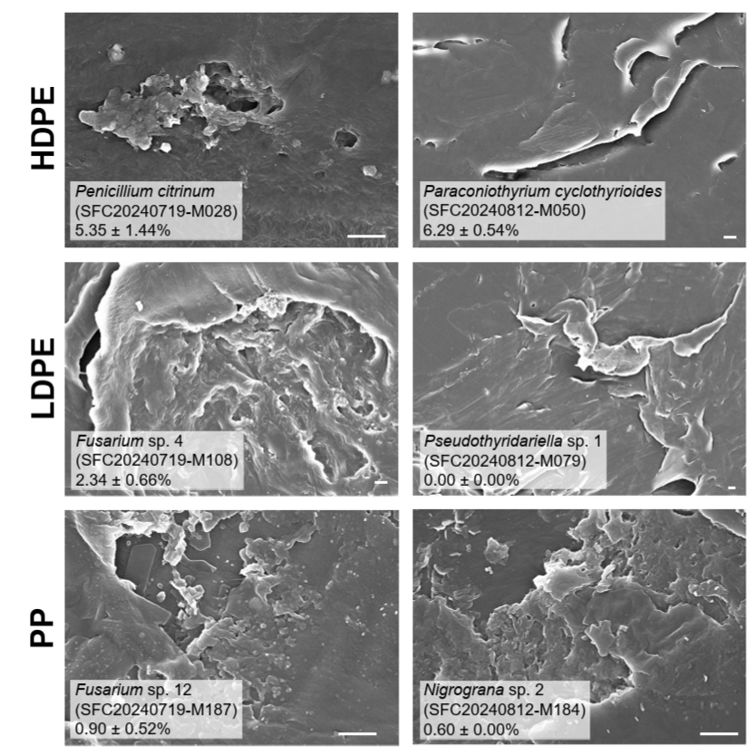
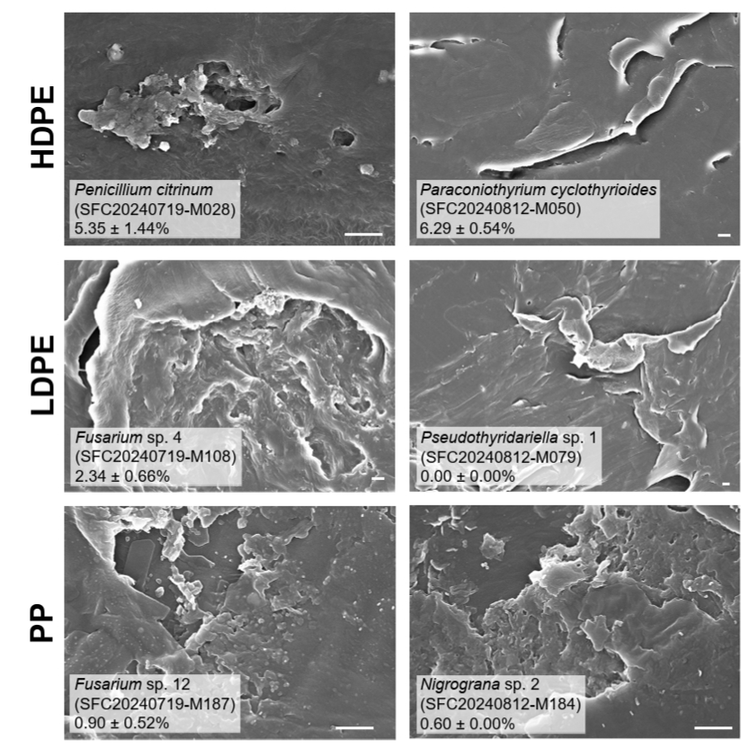
The degradation capacity varied across taxonomic classifications and plastic types (Fig. S1). Most of the genera isolated from HDPE exhibited a substantial plastic degradation capacity. The top-ranking plastic-degrading strains isolated from LDPE were from the genera Cladosporium and Penicillium, whereas some plastic-degrading strains from PP belonged to Aspergillus, Nigrograna, and Penicillium. The stage-level plastic degradation capacity varied across the plastic types and developmental stages (Table S3). SEM observations revealed plastic-specific and stage-dependent surface damage patterns, although visible surface disruption did not always correspond to measurable weight loss (Fig. 6). HDPE disc surfaces showed irregular cavities and fine pores when colonized by early-stage strains, and extensive cracking and lifting when colonized by mature-stage strains. LDPE disc surfaces were altered with substantial erosion, deep indentations, and increased roughness when colonized by early-stage strains, but only surface cracking occurred without apparent weight loss when colonized by mature-stage strains. PP disc surfaces showed alterations by strains from both stages (although to a lesser extent than in the cases of HDPE or LDPE) including cavities with fragmented crystalline material (by early-stage strains) and partial delamination with small pores and plate-like fragments (by mature-stage strains).
Discussion
Temporal progression of plastisphere development in the marine mesocosm
In this study, we investigated the culturable fungal communities in the plastispheres of three common plastic types (HDPE, LDPE, and PP) across two developmental stages (early versus mature) in a marine mesocosm experiment. Despite the relatively short deployment period in seawater, microscopic observations revealed a clear temporal progression in plastisphere development, with sparse microbial attachment in the early stage and a more complex microbial community structure in the mature stage. These trends are consistent with the known marine plastisphere developmental processes (Eich et al., 2015; Jacquin et al., 2021).
Culturable diversity and dominant taxa of fungi colonizing marine plastispheres
Fungi were likewise found to establish dynamically on plastic surfaces during the experimental period. A total of 179 fungal taxa were isolated from the plastic sheets, indicating a substantial fungal diversity associated with marine plastispheres. Notably, a considerable number of taxa were recovered in the early developmental stage, suggesting that fungal colonization occurs rapidly following plastic exposure to marine environments. The genera Penicillium, Cladosporium, Trichoderma, Aspergillus, and Fusarium were dominant across all three plastic types and the two plastisphere developmental stages, which is consistent with previous studies on marine plastisphere-related fungal diversity (Philippe et al., 2023; Rakhmawati et al., 2025). These cosmopolitan saprotrophic fungi exhibit broad environmental tolerance and produce diverse extracellular enzymes (Behera and Das, 2023; Levetin et al., 2016; Ward, 2012), which likely enables their colonization of nutrient-poor plastic surfaces in dynamic marine environments. Cladosporium and Penicillium species were particularly abundant across all plastic types and developmental stages. Several of these species, such as C. rectoides and P. exsudans, have repeatedly been reported across the marine plastisphere (Florio Furno et al., 2025; Kim et al., 2022), suggesting that these genera may play important roles therein.
Temporal shifts in species richness of dominant fungal genera
Although the overall community composition did not differ significantly between developmental stages, temporal shifts in the relative species richnesses of the dominant genera were observed. Cladosporium exhibited higher species richness in the early stage. Several species within this genus are known for their high osmotolerance and capacity to grow under low water activity and oligotrophic conditions (Bensch et al., 2018; Segers et al., 2015; Zalar et al., 2007). These traits may facilitate early colonization when organic resources are limited. By contrast, Trichoderma exhibited a higher relative species richness at the mature stage across all plastic types. Trichoderma species are well-known for producing various hydrolytic and oxidative enzymes, such as laccases and manganese peroxidases (Hoque et al., 2023; Othman et al., 2021; Sowmya et al., 2014). These enzymatic capabilities may be advantageous in the mature stage when microbial biomass and organic matter have accumulated on plastic surfaces.
Environmental drivers shaping fungal community composition
Despite the abovementioned temporal trends, overall fungal community composition did not show statistically significant differences among the developmental stages or plastic types. The lack of substrate-specific clustering may reflect physicochemical properties shared by HDPE, LDPE, and PP, including high hydrophobicity and low surface energy (Encinas et al., 2010). Additionally, the close spatial arrangement of plastic sheets within mesocosm structures likely resulted in relatively homogeneous environmental conditions, reducing substrate-specific selective pressures. Other environmental factors may also have shaped the community compositions. Nutrient concentrations and stoichiometric ratios drive the succession of marine fungal communities in coastal ecosystems (Guo et al., 2015). Moreover, nutrient availability can modulate cross-kingdom interactions between bacteria and fungi, suggesting that such interactions underlie shifts in community composition (Velez et al., 2018). In accordance with this, SEM observations revealed diverse microorganisms colonizing plastic surfaces, together with clear alterations, such as increased roughness, pits, and cavities. Although these features may reflect the combined activity of multiple microbial groups, subsequent weight-loss assays demonstrated that a substantial proportion of the isolated fungal strains (54.6%) exhibited measurable degradation capacities. Furthermore, similar surface damage patterns were reproduced under controlled conditions following fungal inoculation, indicating that fungi contributed to the observed surface modifications. However, as culture-dependent approaches capture only the cultivable fraction of the fungal community, interaction-driven or fine-scale substrate-specific differences may have been underestimated. Interactions among fungi, as well as between fungi and bacteria, are also likely to influence plastisphere assembly and function through processes such as cooperative biofilm formation, nutrient exchange, and metabolic complementarity (Amaral-Zettler et al., 2020; Su et al., 2022; Wang et al., 2021). Because this study focused on culturable fungi and the degradation capacities of individual strains, these potential interactions could not be evaluated directly. Future studies using culture-independent methods, together with co-culture or functional approaches integrating fungal and bacterial community analyses, would provide a more comprehensive understanding of the dynamics, interactions, and functional roles of plastisphere communities.
Plastic type- and stage-dependent variation in fungal degradation capacity
We initially hypothesized that mature-stage strains would exhibit a higher degradation capacity owing to the enrichment of plastic-degrading fungi during later colonization. However, the degradation capacity varied among the plastic types and developmental stages, suggesting that the contribution of fungi to plastic degradation is affected by the physicochemical properties of each plastic sheet. Consistent with our hypothesis, mature-stage strains on HDPE generally showed a higher degradation capacity. This pattern may be consistent with the time-dependent environmental filtering on HDPE surfaces, where prolonged exposure to abiotic weathering (e.g., UV, salinity, and mechanical abrasion) increases surface oxidation and roughness, thereby improving microbial attachment and enzymatic accessibility relative to freshly immersed HDPE (Ghatge et al., 2020; Montazer et al., 2020). By contrast, LDPE showed relatively high degradation by early-stage strains, suggesting that opportunistic early colonizers with high degradation capacity exploit the more accessible physicochemical structure of LDPE, thereby leaving fewer opportunities for later degraders (Mohanan et al., 2020; Pinto et al., 2020). The PP degradation capacity was comparable between strains from each developmental stage, possibly because of the higher crystallinity and hydrophobicity of PP compared to those of HDPE and LDPE, which impede microbial attachment and enzyme access (Choonut et al., 2025; Guo et al., 2024; Srikanth et al., 2022) and may limit degradation regardless of the plastisphere developmental stage.
Taxonomic patterns and functional heterogeneity of plastisphere fungi
Species with high degradation capacities were unevenly distributed across genera; however, some trends in genus-level compositional changes were observed. The strains exhibiting the highest degradation capacity belonged to Aspergillus, Penicillium, Cladosporium, and Trichoderma, which have previously been reported as effective plastic degraders (Kim et al., 2022; Ma et al., 2025; Zeghal et al., 2021). Notably, the observed fungal degradation capacity did not show a strong association with plastic type or developmental stage, suggesting that species-level functional traits are more important determinants of degradation performance than factors depending on the environments from which strains were isolated. However, 45.4% of the strains exhibited no detectable plastic degradation. These non-degrading strains likely fulfilled alternative ecological roles within plastisphere communities. Some may have utilized organic matter derived from other marine organisms, contributing to nutrient cycling rather than direct plastic degradation (Peng et al., 2024; Salazar-Alekseyeva et al., 2023; Wang et al., 2016). Others may have contributed to biofilm formation by producing EPS, thereby enhancing structural stability and facilitating the adhesion of other organisms (Decho and Gutierrez, 2017; Qi et al., 2022). Together, these findings suggest that fungi contribute to plastisphere functioning in diverse ways beyond direct plastic degradation, including biofilm formation, structural stabilization, and nutrient cycling, thereby sustaining the plastisphere as a microecosystem.
Conclusions and Future Perspectives
This study provides novel insights into the composition and functional capacities of culturable fungal communities in the marine plastisphere across different plastic types and developmental stages. Using a cultivation-based approach, we directly assessed plastic degradation at the taxonomic level and found that the degradation potential varied among species, with no consistent relationship between the plastic type or plastisphere developmental stage and degradation capacity. Furthermore, the detection of both degrading and non-degrading fungi indicated the functional heterogeneity of plastisphere communities, suggesting that non-degraders may contribute to other ecological processes, such as nutrient cycling and biofilm structural stability. In addition, the fungal strains identified as active degraders are promising candidates for future applications in plastic bioremediation. Future studies integrating culture-independent approaches with finer temporal sampling over long incubation periods will complement the present findings and further refine our understanding of fungal succession and the ecological role of fungi in marine plastisphere.
Acknowledgments
This research was supported by the Basic Science Research Program of the National Research Foundation of Korea (NRF), funded by the Ministry of Education (Grant number: RS-202400461731), the Management of the Marine and Fishery Bio-resources Center (2026) funded by the National Marine Biodiversity Institute of Korea (MABIK), and Global-Learning & Academic research institution for Master’s‧PhD students, and Postdocs (LAMP) Program of the National Research Foundation of Korea (NRF) grant funded by the Ministry of Education (NO. RS-2024-00444460).
Conflict of Interest
The authors declare that they have no competing interests.
Supplementary Information
The online version contains supplementary material available at https://doi.org/10.71150/jm.2605011
Table S3.
Top plastic-degrading fungal strains identified in this study. Weight-loss (%) values are shown as Means ± standard deviations for each plastic type and exposure time.
jm-2605011-Supplementary-Table-S3.pdf
Fig. S1.
Taxonomic distribution of plastic degradation ability across fungal strains. Heat trees show hierarchical taxonomic relationships and mean weight-loss assay results (%) of fungal strains isolated from (A) HDPE, (B) LDPE, and (C) PP. Each terminal node represents a fungal species, and node size reflects the number of strains within each taxonomic group. Node color intensity represents the mean value of weight-loss assay results (weight loss %).
jm-2605011-Supplementary-Fig-S1.pdf
Fig. 1.Mesocosm experimental design and field photographs of plastic sheets across plastisphere developmental stages. (A) Photograph of the research site where the mesocosm experiment was conducted (top) and schematic illustration of a mesocosm structure (bottom). (B) Photographs of plastic sheets showing surface colonization patterns in the two plastisphere developmental stages; top panels: early stage (seven days after deployment, July 2024); bottom panels: mature stage (30 days after deployment, August 2024).
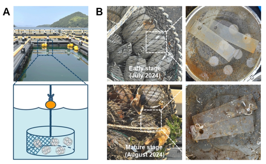
Fig. 2.Scanning electron micrographs of plastic sheet surfaces from the mesocosm experiment. Representative SEM images of high-density polyethylene (HDPE; top), low-density polyethylene (LDPE; middle), and polypropylene (PP; bottom) surfaces sampled at the early (left) and mature (right) stages. Insets in the mature-stage micrographs show higher-magnification views of the regions indicated by arrows, highlighting surface morphology and microbial colonization. Scale bars = 1 μm.
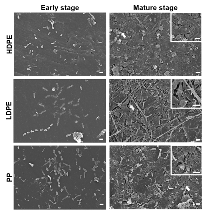
Fig. 3.Culturable fungal communities across plastic types and developmental stages. Treemaps show the relative species richness of fungal genera isolated from high-density polyethylene (HDPE; top), low-density polyethylene (LDPE; middle), and polypropylene (PP; bottom) in the early (left) and mature (right) stages. Box sizes represent the relative number of species within each genus, and numbers indicate the number of species per genus. “Others” represent genera from which only a single species was present; the number indicates the total count of such genera, with individual genus names listed alongside.
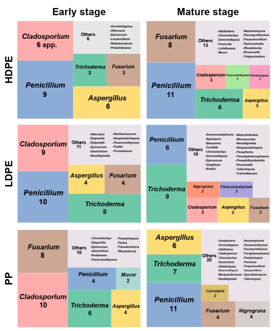
Fig. 4.Alpha and beta diversity of culturable fungal communities across plastic types and developmental stages. (A) Alpha diversity metrics compared among plastic types (high-density polyethylene [HDPE], low-density polyethylene [LDPE], polypropylene [PP]) and developmental stages (early and mature). Top row: inverse Simpson diversity (q = 2); bottom row: exponential Shannon diversity (q = 1). Boxes show medians and interquartile ranges, and reported p- and q-values from statistical tests are indicated. (B) Beta diversity based on Jaccard similarity visualized by principal coordinates analysis. The first (PC1) and second (PC2) principal components explain 28.9% and 20.7% of the variance, respectively. Point indicators are shaped by plastic type and colored by developmental stage.
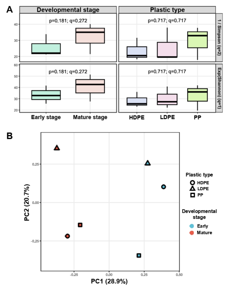
Fig. 5.Degradation performance across isolated fungal strains according to plastic type and developmental origin. Violin plots show the distribution of weight loss percentages after 60 days of incubation with fungal strains tested against the same plastic type from which they were originally isolated. Strains are grouped by the developmental stage of the source plastisphere: early (blue) and mature (pink). Boxplots indicate medians and interquartile ranges. Panels show results for strains from the high-density polyethylene (left), low-density polyethylene (middle), and polypropylene (right) plastispheres.
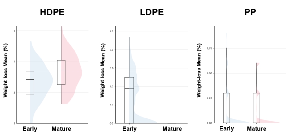
Fig. 6.Surface morphology of plastic discs after 60-day fungal degradation assays. SEM images show plastic surfaces incubated for 60 days with fungal strains exhibiting the highest degradation capacity for each plastisphere type. Left panels: plastics inoculated with strains from the early stage; right panels: plastics inoculated with strains from the mature stage. Top row: high-density polyethylene (HDPE); middle row: low-density polyethylene (LDPE); bottom row: polypropylene (PP). Scale bars = 1 μm.
References
- Amaral-Zettler LA, Zettler ER, Mincer TJ. 2020. Ecology of the plastisphere. Nat Rev Microbiol. 18: 139–151. ArticlePubMedPDF
- Behera AD, Das S. 2023. Ecological insights and potential application of marine filamentous fungi in environmental restoration. Rev Environ Sci Biotechnol. 22: 281–318. ArticlePDF
- Benjamini Y, Hochberg Y. 1995. Controlling the false discovery rate: A practical and powerful approach to multiple testing. J R Stat Soc Ser B Methodol. 57: 289–300. ArticlePDF
- Bensch K, Groenewald JZ, Meijer M, Dijksterhuis J, Jurjević Ž, et al. 2018. Cladosporium species in indoor environments. Stud Mycol. 89: 177–301. Article
- Berger T, Whitner S, Rehar R, Amend AS. 2026. Multi-omics insights into the enzymatic degradation of polyurethane by marine fungi. J Hazard Mater Plast. 2: 100040.Article
- Choonut A, Wongfaed N, Wongthong L, Poolpol A, Chaikitkaew S, et al. 2025. Microbial degradation of polypropylene microplastics and concomitant polyhydroxybutyrate production: An integrated bioremediation approach with metagenomic insights. J Hazard Mater. 490: 137806.Article
- Decho AW, Gutierrez T. 2017. Microbial extracellular polymeric substances (EPSs) in ocean systems. Front Microbiol. 8: 922.ArticlePubMedPMC
- Dixon P. 2003. VEGAN, a package of R functions for community ecology. J Veg Sci. 14: 927–930. ArticleLink
- Dobretsov S, Abed RM, Teplitski M. 2013. Mini-review: Inhibition of biofouling by marine microorganisms. Biofouling. 29: 423–441. ArticlePubMed
- Eich A, Mildenberger T, Laforsch C, Weber M. 2015. Biofilm and diatom succession on polyethylene (PE) and biodegradable plastic bags in two marine habitats: Early signs of degradation in the pelagic and benthic zone? PLoS One. 10: e0137201. ArticlePubMedPMC
- Encinas N, Díaz-Benito B, Abenojar J, Martínez M. 2010. Extreme durability of wettability changes on polyolefin surfaces by atmospheric pressure plasma torch. Surf Coat Technol. 205: 396–402. Article
- Florio Furno M, Laizé V, Arduino I, Pham GN, Spina F, et al. 2025. Bioprospecting marine fungi from the plastisphere: Osteogenic and antiviral activities of fungal extracts. Mar Drugs. 23: 115.ArticlePubMedPMC
- Foster ZS, Sharpton TJ, Grünwald NJ. 2017. Metacoder: An R package for visualization and manipulation of community taxonomic diversity data. PLoS Comput Biol. 13: e1005404. ArticlePubMedPMC
- Friedman M. 1937. The use of ranks to avoid the assumption of normality implicit in the analysis of variance. J Am Stat Assoc. 32: 675–701. Article
- Gao R, Liu R, Sun C. 2022. A marine fungus Alternaria alternata FB1 efficiently degrades polyethylene. J Hazard Mater. 431: 128617.ArticlePubMed
- Geyer R, Jambeck JR, Law KL. 2017. Production, use, and fate of all plastics ever made. Sci Adv. 3: e1700782. ArticlePMC
- Ghatge S, Yang Y, Ahn JH, Hur HG. 2020. Biodegradation of polyethylene: A brief review. Appl Biol Chem. 63: 27.ArticlePDF
- Gower JC. 1966. Some distance properties of latent root and vector methods used in multivariate analysis. Biometrika. 53: 325–338. Article
- Guo RT, Li X, Yang Y, Huang JW, Shen P, et al. 2024. Natural and engineered enzymes for polyester degradation: A review. Environ Chem Lett. 22: 1275–1296. ArticlePDF
- Guo X, Zhang Q, Zhang X, Zhang J, Gong J. 2015. Marine fungal communities in water and surface sediment of a sea cucumber farming system: Habitat-differentiated distribution and nutrients driving succession. Fungal Ecol. 14: 87–98. Article
- Hill MO. 1973. Diversity and evenness: A unifying notation and its consequences. Ecology. 54: 427–432. ArticleLink
- Hoque RA, Yadav M, Yadav HS, Boruah R. 2023. Purification and characterization of novel manganese peroxidase from Trichoderma parestonica and its bio-conversion study of toxic arylamine. Anal Chem Lett. 13: 641–659. Article
- Hurlbert SH. 1971. The nonconcept of species diversity: A critique and alternative parameters. Ecology. 52: 577–586. ArticlePubMedLink
- Jacquin J, Callac N, Cheng J, Giraud C, Gorand Y, et al. 2021. Microbial diversity and activity during the biodegradation in seawater of various substitutes to conventional plastic cotton swab sticks. Front Microbiol. 12: 604395.ArticlePubMedPMC
- Jacquin J, Cheng J, Odobel C, Pandin C, Conan P, et al. 2019. Microbial ecotoxicology of marine plastic debris: A review on colonization and biodegradation by the "plastisphere". Front Microbiol. 10: 865.ArticlePubMedPMC
- Kim SH, Lee JW, Kim JS, Lee W, Park MS, et al. 2022. Plastic-inhabiting fungi in marine environments and PCL degradation activity. Antonie van Leeuwenhoek. 115: 1379–1392. ArticlePubMedPMCPDF
- Lande R. 1996. Statistics and partitioning of species diversity, and similarity among multiple communities. Oikos. 76: 5–13. Article
- Levetin E, Horner WE, Scott JA, Barnes C, Baxi S, et al. 2016. Taxonomy of allergenic fungi. J Allergy Clin Immunol Pract. 4: 375–385.e371. ArticlePubMed
- Li K, Xu L, Bai X, Zhang G, Zhang M, et al. 2024. Potential environmental risks of field bio/non-degradable microplastic from mulching residues in farmland: Evidence from metagenomic analysis of plastisphere. J Hazard Mater. 465: 133428.Article
- Ma Y, Zhou Y, Zheng D, Bu W, Wang F, et al. 2025. Nanoplastics and fungi: Exploring dual roles in degradation and pathogenicity. Front Microbiol. 16: 1679160.ArticlePubMedPMC
- Mathur G, Prasad R. 2012. Degradation of polyurethane by Aspergillus flavus (ITCC 6051) isolated from soil. Appl Biochem Biotechnol. 167: 1595–1602. ArticlePubMedPDF
- Mohanan N, Montazer Z, Sharma PK, Levin DB. 2020. Microbial and enzymatic degradation of synthetic plastics. Front Microbiol. 11: 580709.Article
- Montazer Z, Habibi Najafi MB, Levin DB. 2020. Challenges with verifying microbial degradation of polyethylene. Polymers. 12: 123.ArticlePubMed
- Odobel C, Dussud C, Philip L, Derippe G, Lauters M, et al. 2021. Bacterial abundance, diversity and activity during long-term colonization of non-biodegradable and biodegradable plastics in seawater. Front Microbiol. 12: 734782.ArticlePubMedPMC
- Othman AM, Mahmoud M, Abdelraof M, Karim GSA, Elsayed AM. 2021. Enhancement of laccase production from a newly isolated Trichoderma harzianum S7113 using submerged fermentation: Optimization of production medium via central composite design and its application for hydroquinone degradation. Int J Biol Macromol. 192: 219–231. ArticlePubMed
- Peng X, Amend AS, Baltar F, Blanco-Bercial L, Breyer E, et al. 2024. Planktonic marine fungi: A review. J Geophys Res Biogeosci. 129: e2023JG007887. Article
- Philippe A, Noël C, Eyheraguibel B, Briand JF, Paul-Pont I, et al. 2023. Fungal diversity and dynamics during long-term immersion of conventional and biodegradable plastics in the marine environment. Diversity. 15: 579.Article
- Philippe A, Salaun M, Quemener M, Noël C, Tallec K, et al. 2024. Colonization and biodegradation potential of fungal communities on immersed polystyrene vs. biodegradable plastics: A time series study in a marina environment. J Fungi (Basel). 10: 428.ArticlePubMedPMC
- Pinto M, Polania Zenner P, Langer TM, Harrison J, Simon M, et al. 2020. Putative degraders of low-density polyethylene-derived compounds are ubiquitous members of plastic-associated bacterial communities in the marine environment. Environ Microbiol. 22: 4779–4793. ArticlePubMedPMCLink
- Qi M, Zheng C, Wu W, Yu G, Wang P. 2022. Exopolysaccharides from marine microbes: Source, structure and application. Mar Drugs. 20: 512.ArticlePubMed
- Qian PY, Cheng A, Wang R, Zhang R. 2022. Marine biofilms: Diversity, interactions and biofouling. Nat Rev Microbiol. 20: 671–684. ArticlePubMedPDF
- Rakhmawati A, Octavia B, Aminatun T, Budiasih KS, Ariyanti D. 2025. Mapping the diversity of beach fungal plastisphere: Insights from metagenomic approaches. Trends Sci. 22: 9575.ArticleLink
- Rummel CD, Lechtenfeld OJ, Kallies R, Benke A, Herzsprung P, et al. 2021. Conditioning film and early biofilm succession on plastic surfaces. Environ Sci Technol. 55: 11006–11018. ArticleLink
- Salazar-Alekseyeva K, Herndl GJ, Baltar F. 2023. Release of cell-free enzymes by marine pelagic fungal strains. Front Fungal Biol. 4: 1209265.ArticlePubMed
- Sathiyabama M, Boomija R, Sathiyamoorthy T, Mathivanan N, Balaji R. 2024. Mycodegradation of low-density polyethylene by Cladosporium sphaerospermum, isolated from platisphere. Sci Rep. 14: 8351.ArticlePubMedPMCPDF
- Segers FJ, Meijer M, Houbraken J, Samson RA, Wösten HA, et al. 2015. Xerotolerant Cladosporium sphaerospermum are predominant on indoor surfaces compared to other Cladosporium species. PLoS One. 10: e0145415. ArticlePubMedPMC
- Seo CW, Yoo S, Cho Y, Kim JS, Steinegger M, et al. 2025. FunVIP: Fungal validation and identification pipeline based on phylogenetic analysis. J Microbiol. 63: e2411017. ArticlePubMedPDF
- Sowmya H, Krishnappa M, Thippeswamy B. 2014. Degradation of polyethylene by Trichoderma harzianum—SEM, FTIR, and NMR analyses. Environ Monit Assess. 186: 6577–6586. ArticlePubMedPDF
- Srikanth M, Sandeep T, Sucharitha K, Godi S. 2022. Biodegradation of plastic polymers by fungi: A brief review. Bioresour Bioprocess. 9: 42.ArticlePDF
- Su X, Yang L, Yang K, Tang Y, Wen T, et al. 2022. Estuarine plastisphere as an overlooked source of N2O production. Nat Commun. 13: 3884.ArticlePubMedPDF
- UNEP, United Nations Environment Programme. 2021. From pollution to solution. A global assessment of marine litter and plastic pollution. Available from http://doi.org/10.13140/RG.2.2.33577.31845Article
- Vaksmaa A, Vielfaure H, Polerecky L, Kienhuis M, Van Der Meer M, et al. 2024. Biodegradation of polyethylene by the marine fungus Parengyodontium album. Sci Total Environ. 934: 172819.Article
- Velez P, Espinosa-Asuar L, Figueroa M, Gasca-Pineda J, Aguirre-von-Wobeser E, et al. 2018. Nutrient dependent cross-kingdom interactions: Fungi and bacteria from an oligotrophic desert oasis. Front Microbiol. 9: 1755.Article
- Wang Y, Barth D, Tamminen A, Wiebe MG. 2016. Growth of marine fungi on polymeric substrates. BMC Biotechnol. 16: 3.ArticlePubMed
- Wang P, Song T, Bu J, Zhang Y, Liu J, et al. 2022. Does bacterial community succession within the polyethylene mulching film plastisphere drive biodegradation? Sci Total Environ. 824: 153884.Article
- Wang L, Tong J, Li Y, Zhu J, Zhang W, et al. 2021. Bacterial and fungal assemblages and functions associated with biofilms differ between diverse types of plastic debris in a freshwater system. Environ Res. 196: 110371.ArticlePubMed
- Ward OP. 2012. Production of recombinant proteins by filamentous fungi. Biotechnol Adv. 30: 1119–1139. Article
- Wickham H. 2011. ggplot2. WIREs Comp Stat. 3: 180–185. ArticleLink
- Wilcoxon F. 1945. Individual comparisons by ranking methods. Biometrics. 1: 80–83. Article
- Yu RS, Yang YF, Singh S. 2023. Global analysis of marine plastics and implications of control measure strategies. Front Mar Sci. 10: 1305091.Article
- Zalar P, De Hoog G, Schroers HJ, Crous P, Groenewald J, et al. 2007. Phylogeny and ecology of the ubiquitous saprobe Cladosporium sphaerospermum, with descriptions of seven new species from hypersaline environments. Stud Mycol. 58: 157–183. ArticlePubMedPMC
- Zeghal E, Vaksmaa A, Vielfaure H, Boekhout T, Niemann H. 2021. The potential role of marine fungi in plastic degradation – A review. Front Mar Sci. 8: 738877.Article
- Zettler ER, Mincer TJ, Amaral-Zettler LA. 2013. Life in the "plastisphere": Microbial communities on plastic marine debris. Environ Sci Technol. 47: 7137–7146. Article
- Zhang K, Hu J, Yang S, Xu W, Wang Z, et al. 2022. Biodegradation of polyester polyurethane by the marine fungus Cladosporium halotolerans 6UPA1. J Hazard Mater. 437: 129406.ArticlePubMed
Citations
Citations to this article as recorded by

 MSK
MSK




 ePub Link
 Cite this Article
 XML Download