1Department of Microbiology, Keimyung University School of Medicine, Daegu 42601, Republic of Korea
2Division of Life Science, Department of Bio & Medical Big Data (BK21 Four Program), Research Institute of Life Science, Gyeongsang National University, Jinju 52828, Republic of Korea
3Department of Infectious Diseases, Keimyung University Dongsan Hospital, Keimyung University School of Medicine, Daegu 42601, Republic of Korea
4Center for Bio-imaging & Translational Research, Korea Basic Science Institute, Cheongju 28119, Republic of Korea
5Department of Chemistry, Hanyang University, Seoul 04763, Republic of Korea
6Department of Biological Sciences, University of Ulsan, Ulsan 44610, Republic of Korea
7Basic-Clinic Convergence Research Institute, University of Ulsan, Ulsan 44610, Republic of Korea
This is an Open Access article distributed under the terms of the Creative Commons Attribution 4.0 International License (CC BY 4.0) (https://creativecommons.org/licenses/by/4.0/) which permits unrestricted use, distribution, and reproduction in any medium, provided the original work is properly cited.
Hypervirulent Klebsiella pneumoniae (hvKp) is an emerging pathogen that causes severe community-acquired infections; however, the immune mechanisms controlling intracellular hvKp have yet to be clearly defined. In this study, we investigated the therapeutic potential of berberine against hvKp infection and elucidated the underlying molecular mechanisms using macrophage cell models and in vivo zebrafish models. Berberine significantly reduced intracellular hvKp survival in macrophages and improved survival in hvKp-infected zebrafish. Berberine markedly attenuated proinflammatory cytokine production and inhibited the c-Jun N-terminal kinase (JNK) and extracellular signal-regulated kinase (ERK) signaling pathways. Notably, we found that hvKp exploited host lipid droplets (LD) biosynthesis to support its intracellular survival, and berberine effectively suppressed LD accumulation. Mechanistically, berberine promoted the nuclear translocation of transcription factor EB (TFEB), thereby enhancing lipolysis. Although berberine upregulated autophagy-related gene expression during hvKp infection, it did not induce lipophagy, the selective autophagic degradation of LD. Collectively, these findings indicate that berberine has potential as a therapeutic agent against hvKp infection by modulating host lipid metabolism to restrict bacterial intracellular survival.
Klebsiella pneumoniae (Kp) is an opportunistic Gram-negative bacterium belonging to the Enterobacteriaceae family that primarily colonizes the human gastrointestinal tract and nasopharynx. It is also widely found on the mucosal surfaces of animals and in natural environments, such as water and soil (Wang et al., 2020). In immunocompromised individuals, it can invade the host and cause a wide range of severe infections, including pneumonia, urinary tract infections, and sepsis (Abbas et al., 2024). The increase in multidrug-resistant Kp strains has become a critical public health concern, leading to high morbidity and mortality worldwide (Mohd Asri et al., 2021). Among these strains, hypervirulent Kp (hvKp) has emerged as a clinically significant pathogen with higher virulence than classical Kp (cKp) (Russo and Marr, 2019). HvKp is characterized by a hypermucoviscous polysaccharide capsule that helps the bacterium to evade host immune defenses (Liu et al., 2025; Xu et al., 2021) and allows it to cause invasive infections, even in healthy individuals. Furthermore, hvKp strains that acquire antibiotic resistance pose a serious threat to public health because of their enhanced ability to spread in both community and healthcare settings (Marr and Russo, 2019). Considering the clinical importance of hvKp, a comprehensive understanding of host defense mechanisms against this pathogen is vital for developing effective therapeutic strategies.
Berberine is a plant-derived isoquinoline alkaloid with a long history of use in traditional medicines (Neag et al., 2018). Recent studies have highlighted its broad pharmacological effects, including anti-inflammatory, antidiabetic, and lipid-regulatory properties (Cai et al., 2023; Neag et al., 2018). In macrophages, berberine has been shown to modulate lipid metabolism, which plays a critical role in the maintenance of homeostasis and host defense (Li et al., 2017). This modulation is particularly relevant in the context of hvKp infections, in which macrophage function is compromised. Therefore, understanding how berberine influences lipid metabolism in macrophages during hvKp infection may provide insights into potential therapeutic strategies.
Lipid droplets (LD) are intracellular organelles that store neutral lipids, such as triacylglycerols (TG) and cholesterol esters, surrounded by a phospholipid monolayer embedded with LD-associated proteins (Olzmann and Carvalho, 2019). Proper regulation of LD content is essential for maintaining cellular homeostasis, as excessive LD accumulation leads to lipotoxicity, organelle dysfunction, and the development of metabolic diseases such as non-alcoholic fatty liver disease and obesity (Mashek, 2021). To prevent such deleterious consequences, cells have evolved tightly regulated mechanisms to degrade LD and release stored fatty acids for energy production or membrane biosynthesis (Zechner et al., 2012). The degradation of LD occurs through two main mechanisms: lipophagy and lipolysis (Corbo and Chung, 2024; Singh et al., 2009; Zechner et al., 2017). Lipophagy is a selective form of autophagy that targets LD for lysosomal degradation and plays a crucial role in cellular metabolism, energy homeostasis, and protection against lipotoxicity (Kounakis et al., 2019). Lipolysis is the enzymatic breakdown of LD into free fatty acids and glycerol, which provide energy and metabolic intermediates for cellular processes (Bolsoni-Lopes and Alonso-Vale, 2015). In immune cells, such as macrophages, lipolysis plays an important role not only in energy homeostasis but also in modulating immune responses and pathogen defense (Leopold Wager et al., 2019; Wang et al., 2019). Recent studies have suggested that the activation of lipolysis can restrict the growth of certain pathogens by depriving them of lipid resources (Vrieling et al., 2019). Therefore, defining how lipolysis is regulated in infected macrophages may clarify whether host lipid metabolism can be therapeutically targeted during hvKp infection. This study aimed to determine whether berberine limits hvKp survival by modulating macrophage inflammatory responses and lipid metabolism, with particular attention to LD turnover, transcription factor EB (TFEB) signaling, autophagy, and lipolysis.
Materials and Methods
Cell culture
For all experiments, RAW 264.7 cells (TIB-71, American Type Culture Collection) were maintained at 37℃ in 5% CO2 incubator using Dulbecco’s Modified Eagle’s Medium (DMEM; Welgene, Korea) containing 10% fetal bovine serum (Welgene) and 1% antibiotics (Gibco BRL, USA).
Bacterial strains and culture
Samples obtained from patients diagnosed with liver abscess or pneumonia at Keimyung University Dongsan Hospital (Korea) were used to isolate Kp strains, DSMC-K210, DSMC-K285, and DSMC-K305. Capsular serotypes and virulence-associated genes were analyzed by polymerase chain reaction (PCR). Identification of the isolates and antimicrobial susceptibility profiles were assessed using the VITEK 2 automated system (bioMérieux, France). In addition, extended-spectrum β-lactamase production was determined according to Clinical and Laboratory Standards Institute guidelines using an agar dilution method with cefotaxime, ceftazidime, and ceftazidime-clavulanate. The clinical Kp strains used in this study are listed in Table S1. For preservation, bacteria were stored at –80℃ after addition of 15% (v/v) glycerol (Sigma-Aldrich, USA). To prepare inocula for infection experiments, frozen bacterial stocks were thawed, and Kp strains were cultured overnight in Luria-Bertani (LB) broth at 37℃, followed by subculture in fresh medium until reaching an optical density of approximately 0.1 at 600 nm. Before infection assays, viable counts were determined by measuring colony-forming units (CFUs).
Infection and treatments
RAW 264.7 cells were seeded into 24-well plates at a density of 2 × 10⁵ cells/well and infected with the indicated Kp strains at a multiplicity of infection (MOI) of 1 or 40 for 1 h. Cells were then washed three times with phosphate-buffered saline (PBS). To eliminate extracellular bacteria, cells were incubated in fresh medium containing 100 μg/ml gentamicin (15710-072, Gibco) for 30 min. The medium was replaced with fresh medium, and berberine (B3251, Sigma-Aldrich) was added following gentamicin treatment and maintained throughout the experiment. To inhibit lipolysis, cells were treated with atglistatin (15284, Cayman Chemical, USA) for 1 h prior to infection, and treatment was maintained throughout the infection period. All cells were incubated at 37°C in 5% CO₂. All treatments were dissolved in dimethyl sulfoxide (DMSO; 20–139, Sigma-Aldrich).
Cell viability assay
RAW 264.7 cells were plated in 96-well plates at a density of 4 × 10⁴ cells/well and allowed to adhere for 18 h at 37℃ in 5% CO2 incubator. Following the treatment with berberine for 6 h, MTS reagent of the CellTiter 96® AQueous One Solution Cell Proliferation Assay kit (MTS; G3580, Promega, USA) was then added to each well and incubated for 30 min at 37℃. Absorbance was recorded at 490 nm using a Synergy/HTX multimode microplate reader (BioTek Instruments, Inc., USA). Relative cell viability was determined against untreated controls set at 100% from three independent experiments performed in triplicate.
RNA isolation and real-time quantitative PCR (RT-qPCR)
Total RNA was isolated using TRIzol reagent (15596-026, Thermo Fisher Scientific, USA). Complementary DNA (cDNA) was synthesized from the extracted RNA using a reverse transcriptase premix (EBT-1515, Elpis Biotech, Korea). Gene expression analysis by qPCR was conducted using the qTOWER3 PCR thermal cycler (Analytik Jena, Germany) in combination with THUNDERBIRD SYBR qPCR mix (QPS-201, TOYOBO, Japan), and relative expression levels were calculated using the 2-ΔΔCt method and normalized to β-actin or Gapdh. The mouse primer sequences used in this study are listed in Table S2.
CFU assay in macrophages
RAW 264.7 cells were infected with hvKp (DSMC-K210 or DSMC-K285, MOI of 40) or cKp (DSMC-K305, MOI of 1) for 1 h at 37℃ in a 5% CO2 atmosphere using antibiotic-free medium. To eliminate extracellular bacteria, infected cells were exposed to medium containing 100 µg/ml gentamicin for 30 min after washing with PBS. The cells were then washed and cultured in fresh medium for the indicated time. For intracellular bacterial quantification, cells were lysed with 1% saponin. Following 10-fold serial dilution, the lysates were spread onto LB agar plates, and CFUs were determined after overnight incubation at 37℃.
Immunoblotting
Cell lysates were prepared using RIPA lysis buffer (89900, Thermo Fisher Scientific), and the lysates were clarified by centrifugation to collect the supernatant fractions. A bicinchoninic acid assay kit (23225, Pierce, USA) was used to determine protein concentrations. After loading equal amounts of protein (50 µg/lane), electrophoresis was performed on sodium dodecyl-sulfate polyacrylamide gels (8–12%), and the separated proteins were transferred to nitrocellulose membranes (GE Healthcare Life Science, USA). Membranes were blocked and incubated with primary antibodies listed in Table S3 overnight at 4℃. After washing, membranes were exposed with appropriate secondary antibodies including anti-rabbit IgG (111-035-045) and anti-mouse lgG (115-035-062) (both diluted 1:5,000; Jackson ImmunoResearch Laboratories, USA). Protein signals were visualized using the Immobilon Western Chemiluminescent HRP Substrate (EMD Millipore, Germany) and captured with a ChemiDoc imaging system (Analytik Jena, Germany).
Nuclear and cytoplasmic fractionation
RAW 264.7 cells were infected with hvKp (DSMC-K285, MOI 40) and treated with berberine (50 μM) for the indicated times. Cells were harvested and separated into cytoplasmic and nuclear fractions using NE-PERTM Nuclear and Cytoplasmic Extraction Reagents (78833, Thermo ScientificTM, USA) according to the manufacturer’s instructions. Equal amounts of protein from each fraction were separated by sodium dodecyl-sulfate polyacrylamide gel electrophoresis and subjected to immunoblotting as described above. TFEB levels were analyzed in both the cytoplasmic and nuclear fractions. ACTB and lamin B1 were used as cytoplasmic and nuclear fraction markers, respectively. The nuclear translocation of TFEB was quantified by calculating the ratio of nuclear TFEB (normalized to lamin B1) to cytoplasmic TFEB (normalized to ACTB).
Enzyme-linked immunosorbent assay (ELISA)
RAW 264.7 cells were plated in 24-well plates at a density of 2 × 10⁵ cells/well. Culture supernatants were harvested at 4 h and 6 h after incubation and clarified by centrifugation at 300 × g for 5 min. Levels of tumor necrosis factor-alpha (TNF-α; MTA00B-1) and interleukin-6 (IL-6; M6000B-1) were quantified using mouse Quantikine ELISA kits (R&D Systems, USA).
Immunofluorescence and analysis
The cells grown on coverslips were infected with hvKp and subsequently fixed for 15 min with 4% paraformaldehyde. After fixation, cells were permeabilized for 10 min at room temperature with 0.25% Triton X-100 (Sigma-Aldrich). Samples were then incubated with BODIPY 493/503 (D3922, Molecular Probes, USA), TFEB (BETHYL, Invitrogen), or anti-LC3 antibody (PM036, MBL International) according to the manufacturers’ instructions. Nuclei were counterstained with 4′,6-diamidino-2-phenylindole (DAPI; D9542, Sigma-Aldrich) for 5 min, fluorescence images were captured using a confocal laser-scanning microscope (TCS SP8, Leica Microsystems, Germany) and analyzed using ImageJ software (National Institutes of Health, USA).
Flow cytometric analysis of LC3
To assess LC3 levels, cells were first fixed for 15 min using 4% paraformaldehyde, followed by permeabilization for 10 min with 0.25% Triton X-100 (Sigma-Aldrich). Cells were then blocked and incubated with an anti-LC3 primary antibody (1:100; #83506, Cell Signaling Technology) for 1 h at room temperature. After washing with PBS, cells were incubated for 1 h at room temperature with Alexa Fluor Plus 488 conjugated goat anti-mouse IgG (H+L) secondary antibody (1:400; A11001, Invitrogen, USA). Nuclei were stained with 4′,6-diamidino-2-phenylindole (DAPI; D9542, Sigma-Aldrich) for 5 min at room temperature. LC3-associated fluorescence signals were subsequently analyzed using BD FACScanTM flow cytometer (BD Biosciences, USA), and mean fluorescence intensity values were calculated using FlowJo software (BD Biosciences, USA).
TG level measurements
Intracellular TG levels were quantified using a triglyceride quantification kit (DG-TGC100, DoGenBio, Korea). Cell lysates (50 μl) obtained from hvKp-infected RAW 264.7 cells were transferred to a 96-well plate and mixed with 50 μl of TG reaction solution. The mixture was incubated at room temperature for 30 min in the dark. TG levels were then determined through measurement of absorbance at 570 nm using a Synergy/HTX multimode microplate reader (BioTek Instruments, Inc.).
Transmission electron microscopy
To perform transmission electron microscopy analysis, the samples were fixed on ice for 2 h in 2.5% glutaraldehyde and post-fixed with 1% osmium tetroxide. After washing with PBS, the specimens were dehydrated through graded ethanol and propylene oxide solutions prior to Epon 812 embedding and polymerization at 60°C for 24 h. Sections of 70 nm thickness were mounted on 75-mesh copper grids and double-stained with 4% uranyl acetate and lead citrate. Imaging was conducted using a KBSI Bio-HVEM system (JEM-1400Plus at 120 kV and JEM-1000BEF at 1000 kV; JEOL, Japan).
Free glycerol quantification
To quantify free glycerol, cell culture supernatants were first centrifuged at 3,000 rpm for 3 min, and the clarified fractions were subsequently collected. These samples were then reacted with reagents from the Free Glycerol Kit (ab65337, Abcam, UK) in a 96-well plate for 30 min at room temperature in the dark. The absorbance at 570 nm was measured using a Synergy/HTX multimode microplate reader (BioTek Instruments, Inc.).
Zebrafish (ZF) husbandry
Wild-type Danio rerio (AB strain) was provided by the Korea Zebrafish Resource Center. The fish were maintained in an in-house ZF facility under a 14:10 h light/dark cycle. They received brine shrimp twice daily, with an additional feeding on the day before mating. After natural spawning, the embryos were collected in fish water prepared by dissolving 1 g of Instant Ocean sea salt in 1 L of dH2O, and the pH was adjusted to 7.0 using NaHCO3 (Takaki et al., 2013). The fertilized eggs were raised in Petri dishes for up to 3 days post-fertilization (dpf), with daily water changes. All ZF experiments were conducted under approval of the Animal Research Ethics Committee of Gyeongsang National University (Approval No. GNU-190325-E0014).
Preparation of bacterial inoculum
The infection stock of the hvKp strain expressing green fluorescent protein (GFP) was prepared as previously described (Nguyen et al., 2023). Briefly, the strain was inoculated into LB broth supplemented with kanamycin (50 µg/ml) and cultured to mid-logarithmic phase (OD600 ≈ 0.8). The cell pellet was harvested by centrifugation at 4,000 rpm for 5 min and washed twice with LB broth. To ensure a uniform suspension, the bacterial cells were passed through a 26-gauge needle and divided into 200 µl aliquots for storage at –80°C. To determine a uniform stock concentration, three random tubes were selected, and serial dilutions (10-fold) were plated on LB agar plates containing kanamycin (50 µg/ml). Before injection, the frozen aliquot was thawed, washed twice, and resuspended in PBS to achieve the desired concentration (Marcoleta et al., 2018).
Microinjection of hvKp in ZF larvae through the caudal vein
ZF embryos were dechorionated between 30–48 h post-fertilization. Further, tricaine stock solution was prepared by dissolving 0.4 g of tricaine in 100 ml dH2O with pH adjusted to 7.0 using NaHCO3 and stored at 4℃. Prior to use, 630 µl of tricaine stock was diluted in 10 ml of sterilized fish water, yielding a final concentration of approximately 0.24–0.25 mg/ml (Takaki et al., 2013). ZF larvae were anesthetized and mounted on 1.7% agarose prepared with sterilized fish water. A small volume of tricaine solution was added to the plate to maintain anesthesia throughout the injection. Based on prior optimization, an inoculum of ~5,000 CFU/larva was sufficient to establish infection. Accordingly, ~3 nl of the bacterial suspension (5,000 CFU) was injected via the caudal vein using a Tritech Research Digital microinjector (MINJ-D, Tritech Research, USA). Control larvae were injected with an equivalent volume of sterile PBS to exclude nonspecific effects (Ye et al., 2025).
The injection needles were replaced every 100 larvae or 1 h, whichever occurred first. Consistency of the injection was checked by injecting into sterile PBS and plating on LB agar containing 50 µg/ml kanamycin. Post-injection, larvae were transferred to 96-well plates (2 fish/well) and exposed to the drug solutions (Berberine at 3 or 6 µM). Amikacin (AMK; 25 µM) was used as a positive control. A non-infected DMSO-only group was included separately as a vehicle control to monitor possible DMSO toxicity. The drug solution and fish water were changed daily.
Drug efficacy assessment in hvKp-infected ZF
The in vivo efficacy of berberine was determined by hvKp proliferation (GFP fluorescence intensity within the ZF), larval survival, and bacterial burden after homogenization of ZF larvae. Bacterial proliferation was monitored daily by tracking GFP fluorescence in the infected larvae using the ImageXpress Pico Automated Cell Imaging System (Molecular Devices). The end-point GFP signal intensity was used for comparative analysis. Ten larvae from each treatment group were randomly selected and individually lysed in 2% Triton X-100 in PBS with Tween-20 (Jeon et al., 2022). Samples were homogenized using a handheld homogenizer (D1,000; Benchmark), serially diluted (10-fold), and plated on LB agar containing kanamycin (50 µg/ml). Colonies were counted after an overnight incubation. Survival was monitored daily, and the number of dead larvae in each treatment group was recorded for survival curve analysis.
Statistical analysis
The Mean ± standard deviation was used to present the data obtained from three independent experiments. To evaluate statistical differences, we used Student’s t-test or analysis of variance, depending on the nature of the comparison. GraphPad Prism 7.0 for Windows (GraphPad Software, Inc., USA) was used for all statistical analyses and figure generation, and significance was assigned at different thresholds, including P values below 0.05, 0.005, and 0.001.
Results
Berberine suppresses intracellular survival of hvKp
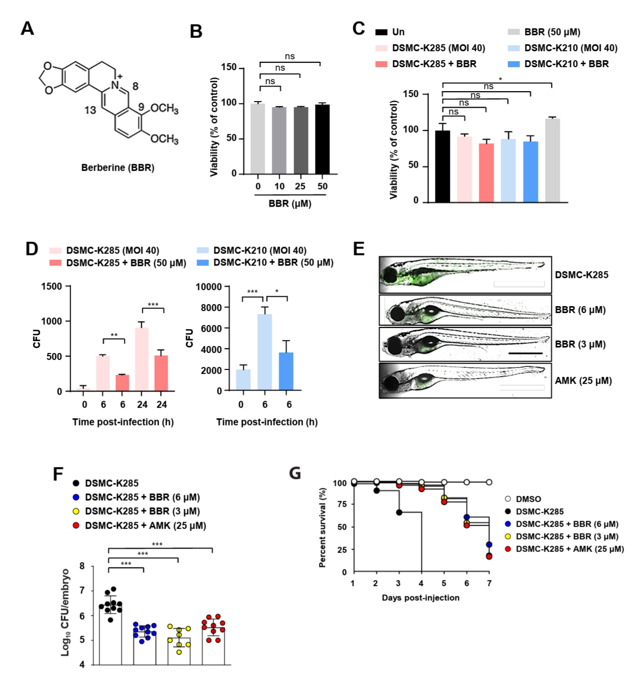
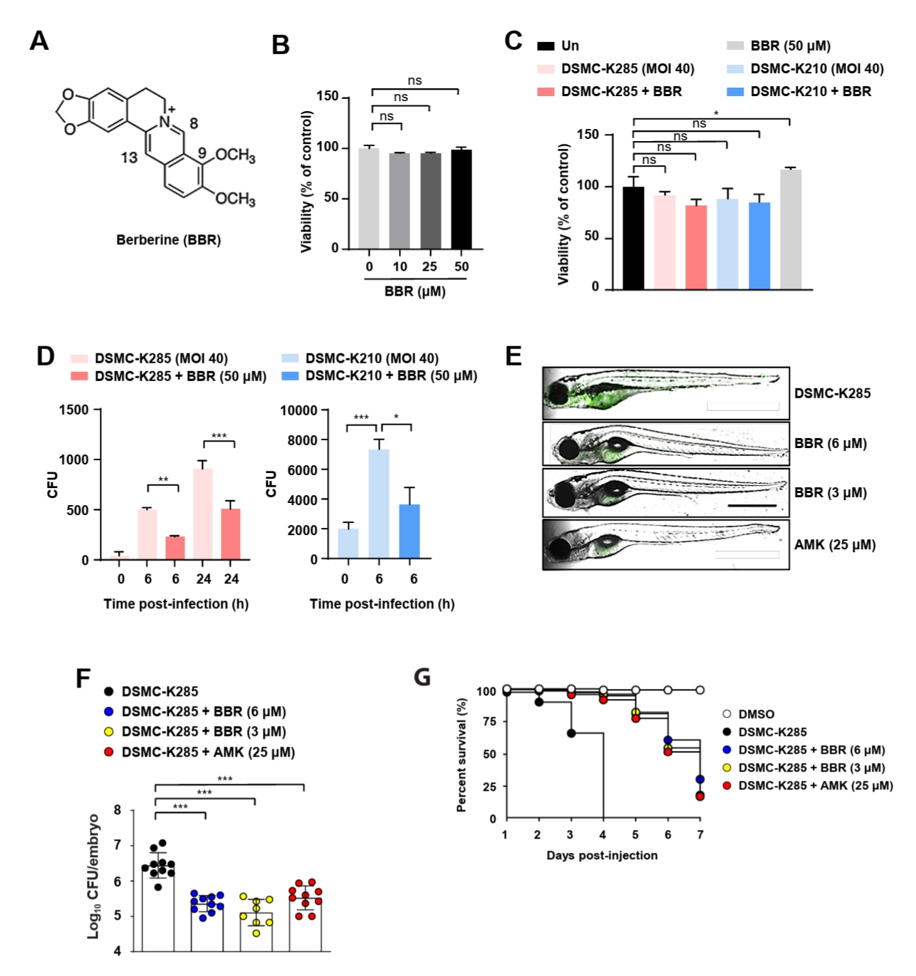
Berberine, an isoquinoline alkaloid (Fig. 1A), regulates macrophage activation and promotes autophagy (Li et al., 2022; Zheng et al., 2021). To evaluate its cytotoxicity prior to functional assays, RAW 264.7 cells were treated with berberine and cell viability was assessed. Berberine treatment did not significantly affect macrophage viability compared with the untreated control (Fig. 1B). To determine whether berberine directly affects bacterial growth, we used two hvKp strains (DSMC-K210 or DSMC-K285) isolated from patients (Jung et al., 2024). Co-treatment with berberine and hvKp did not affect cell viability, indicating that berberine is non-toxic, even during infection (Fig. 1C). Berberine did not affect bacterial growth under these conditions, as no significant differences were observed between the groups (Fig. S1). To evaluate the effects of berberine on intracellular bacterial survival, murine macrophages were infected with hvKp and treated with berberine. A significant reduction in the intracellular bacterial load was observed in the berberine-treated group compared with that in the untreated control group (Fig. 1D). To evaluate the antibacterial effects of berberine in vivo, ZF larvae were infected with GFP-expressing hvKp and treated with berberine. Berberine treatment markedly decreased the GFP fluorescence intensity of hvKp in ZF larvae compared with that in the untreated group, indicating a decreased bacterial burden (Fig. 1E). Berberine treatment significantly reduced bacterial CFU counts in infected ZF embryos (Fig. 1F). Moreover, ZF survival analysis demonstrated that berberine treatment markedly improved host survival compared with that of the infection-only group (Fig. 1G). Similar to the positive control AMK, berberine exhibited protective effects against hvKp infection in vivo. These findings indicate that berberine suppresses intracellular hvKp survival both in vitro and in vivo. Given that berberine did not directly inhibit bacterial growth (Fig. S1) but significantly reduced the intracellular bacterial load in infected macrophages (Fig. 1D), these data suggest that berberine acts through host cell-associated mechanisms rather than through direct antibacterial activity.
Berberine attenuates hvKp-induced inflammatory responses and mitogen-activated protein kinase signaling
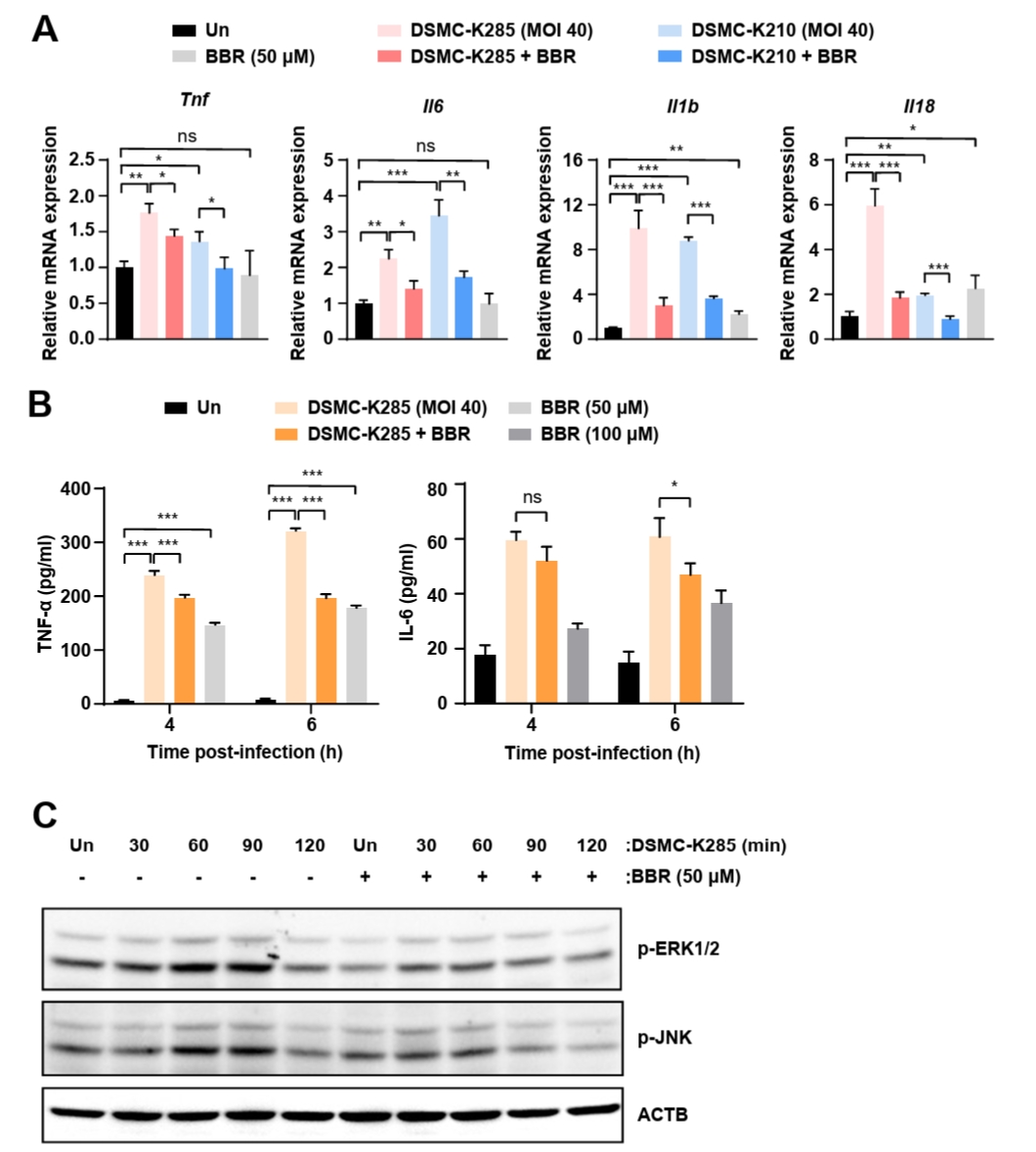
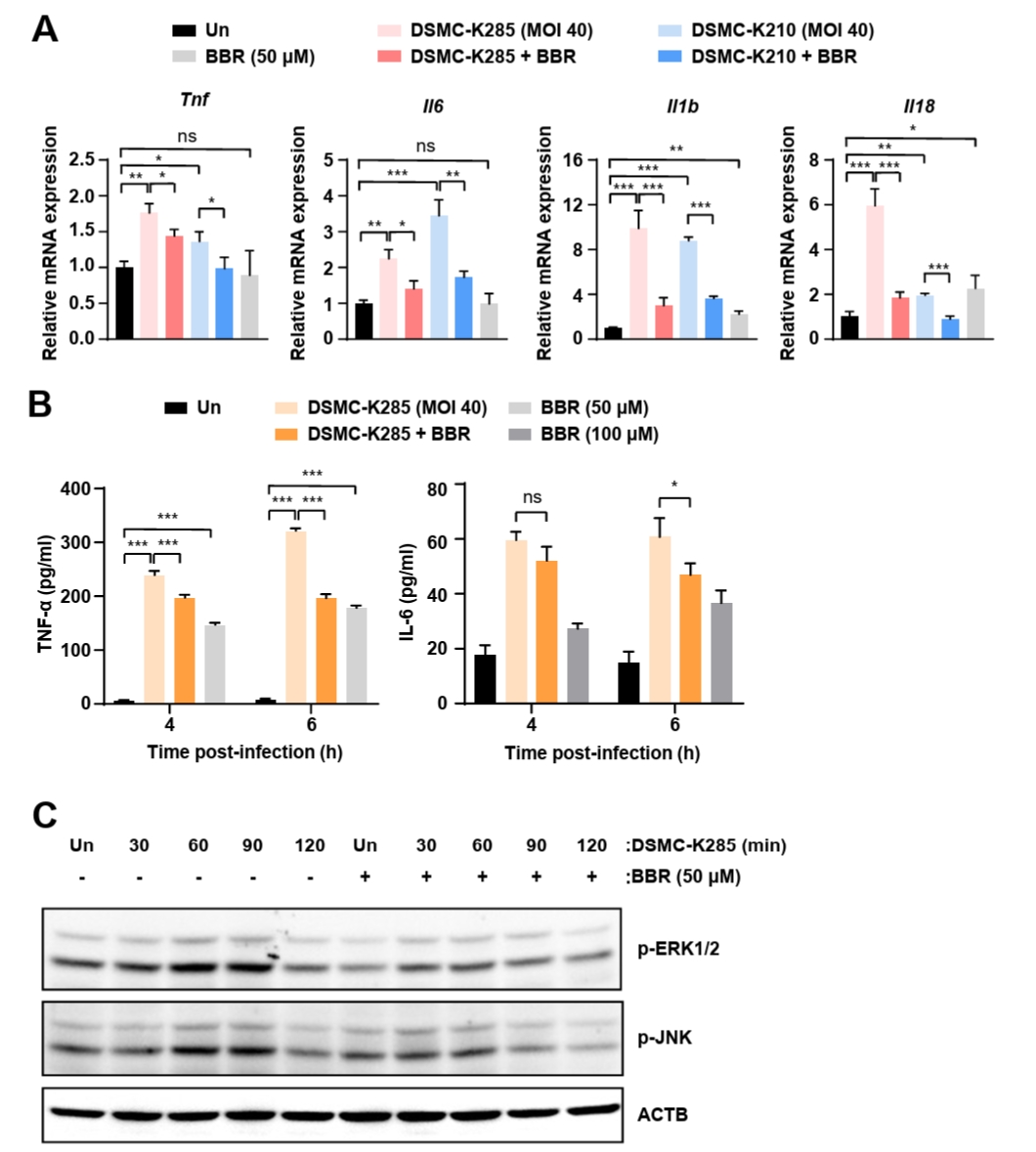
Berberine is known to exert anti-inflammatory effects on macrophages (Zhang et al., 2023). To determine whether berberine similarly attenuates inflammatory responses during hvKp infection, murine macrophages were infected with hvKp strains and treated with berberine. Berberine significantly reduced the mRNA levels of inflammatory cytokines, including interleukin (Il)6, tumor necrosis factor (Tnf)α, Il1b, and Il18, which were elevated following hvKp infection (Fig. 2A). Berberine treatment markedly decreased the secretion of IL-6 and TNF-α in the supernatants of hvKp-infected macrophages (Fig. 2B). In macrophages, inflammatory cytokine expression is regulated by the activation of mitogen-activated protein kinase (MAPK) pathway proteins, including extracellular signal-regulated kinase (ERK) and c-Jun N-terminal kinase (JNK) (Tian et al., 2019). As Kp infection in macrophages is known to increase the phosphorylation of the MAPK pathway proteins ERK and JNK (March et al., 2011; Wu et al., 2006; Xu et al., 2017), we next examined whether hvKp infection induces ERK and JNK phosphorylation and whether berberine co-treatment affects this process. We found that hvKp infection markedly increased ERK and JNK phosphorylation, and that this increase was significantly attenuated by berberine co-treatment (Fig. 2C). These findings indicate that berberine effectively suppresses hvKp-induced inflammatory responses and ERK/JNK phosphorylation in macrophages.
Berberine inhibits LD formation in hvKp-infected murine macrophages
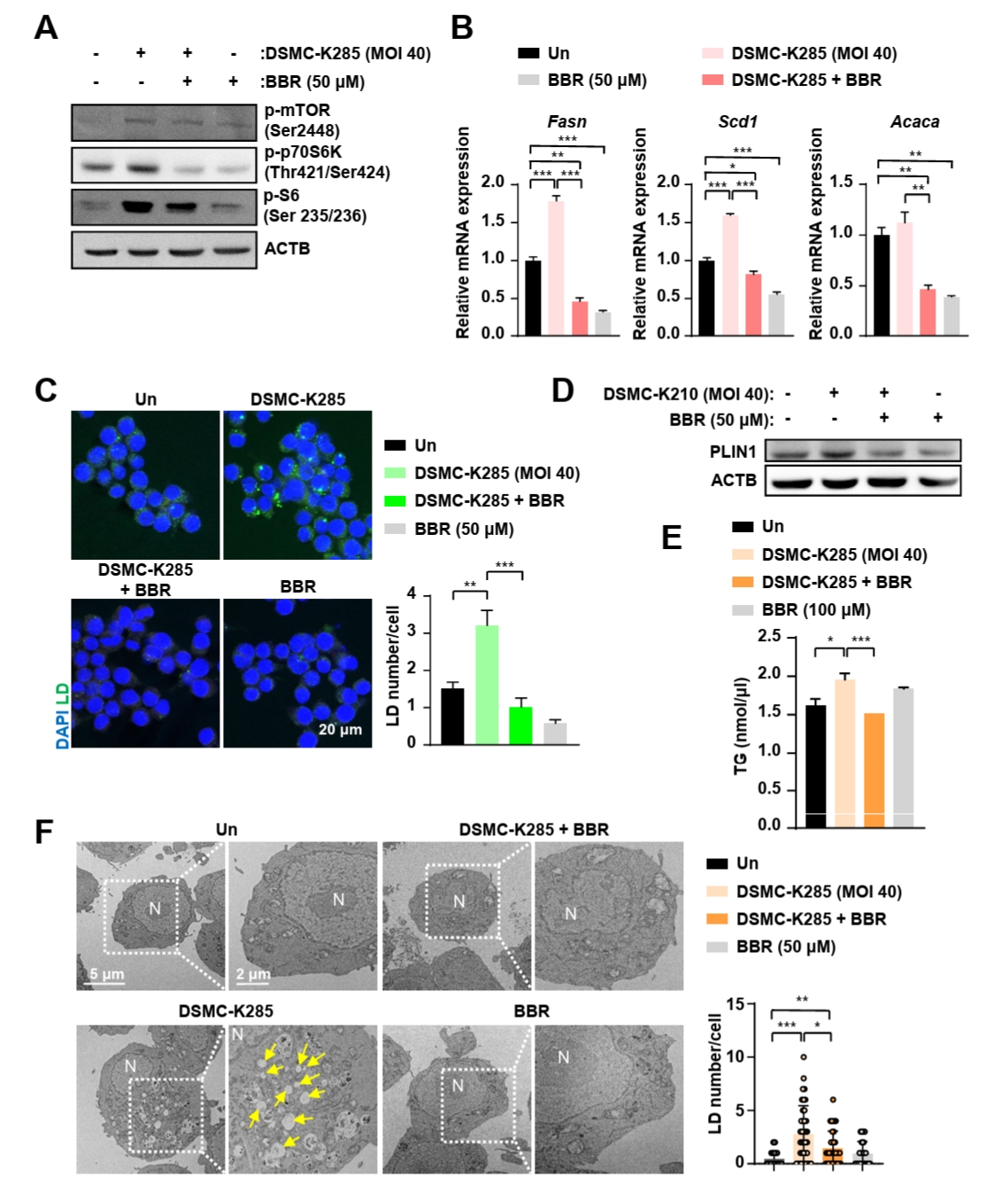
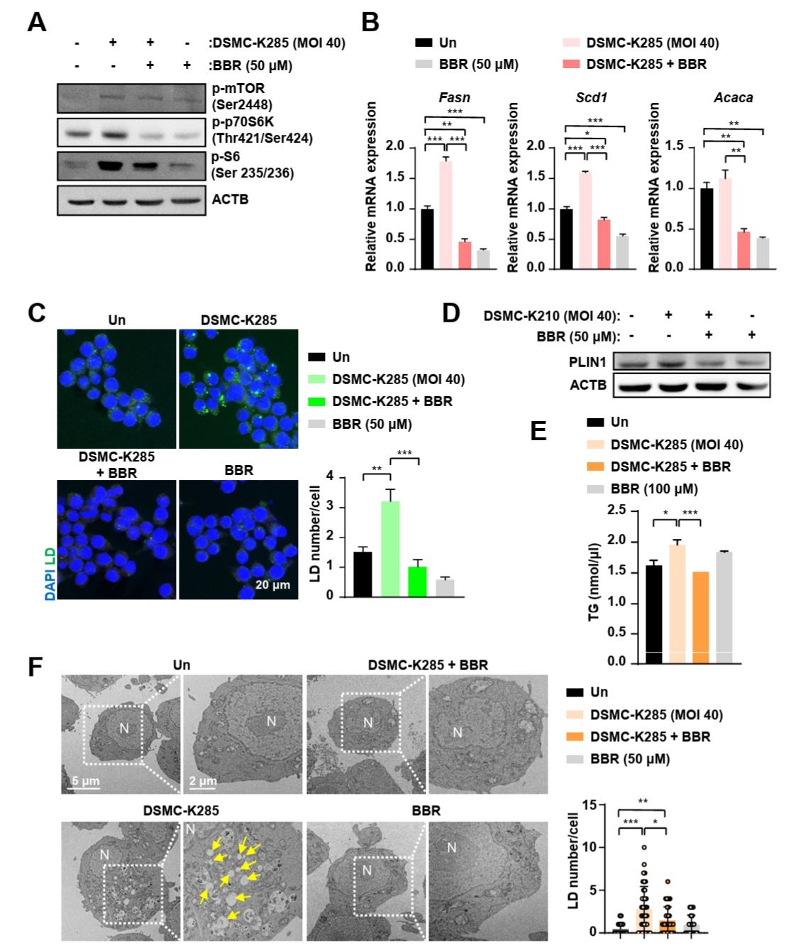
Previous studies have shown that LD formation is crucial for the survival of hvKp within macrophages (Jung et al., 2024). Given that the mammalian target of rapamycin (mTOR) signaling regulates lipid metabolism and contributes to macrophage host defense against Kp infection (Han and Wang, 2018; Jung et al., 2024), we investigated whether berberine modulates LD formation during hvKp infection via the mTOR pathway (Fig. 3A). Infection with hvKp significantly increased the phosphorylation of mTOR and its downstream effectors p70S6K and S6 compared with the uninfected control group. Berberine treatment markedly suppressed hvKp-induced phosphorylation of mTOR, p70S6K, and S6, suggesting that berberine inhibits the mTOR/p70S6K/S6 signaling cascade activated by hvKp infection. As mTOR signaling is known to promote lipogenic gene expression (Düvel et al., 2010), we next assessed the expression of key lipogenesis-related genes, including fatty acid synthase (Fasn), stearoyl-CoA desaturase 1 (Scd1), and acetyl-CoA carboxylase alpha (Acaca). The mRNA levels of these genes were upregulated following hvKp infection, but were markedly reduced by berberine treatment (Fig. 3B). Berberine treatment significantly reduced hvKp-induced LD accumulation, as visualized by confocal microscopy (Fig. 3C). To investigate the changes in LD during hvKp infection and berberine treatment, we examined the expression of perilipin 1 (PLIN1), a major protein localized on the LD membrane. Berberine significantly attenuated the infection-induced upregulation of PLIN1 (Fig. 3D), further supporting that berberine suppresses hvKp-induced LD formation. Furthermore, the quantification of intracellular TG levels demonstrated a marked increase following hvKp infection, which was significantly reduced by berberine co-treatment (Fig. 3E). Transmission electron microscopy further confirmed that hvKp-induced LD formation was attenuated by berberine treatment, validating our observations at the ultrastructural level (Fig. 3F). Collectively, these results demonstrate that berberine attenuates hvKp-induced lipogenesis and LD accumulation in macrophages, at least in part through inhibition of the mTOR/p70S6K/S6 signaling pathway.
Berberine promotes activation and nuclear translocation of transcription factor EB during hvKp infection
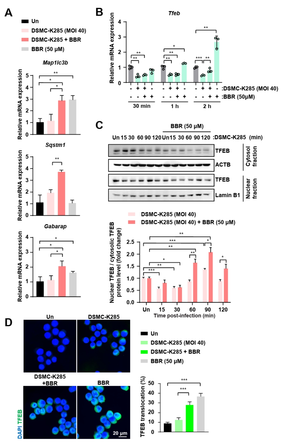
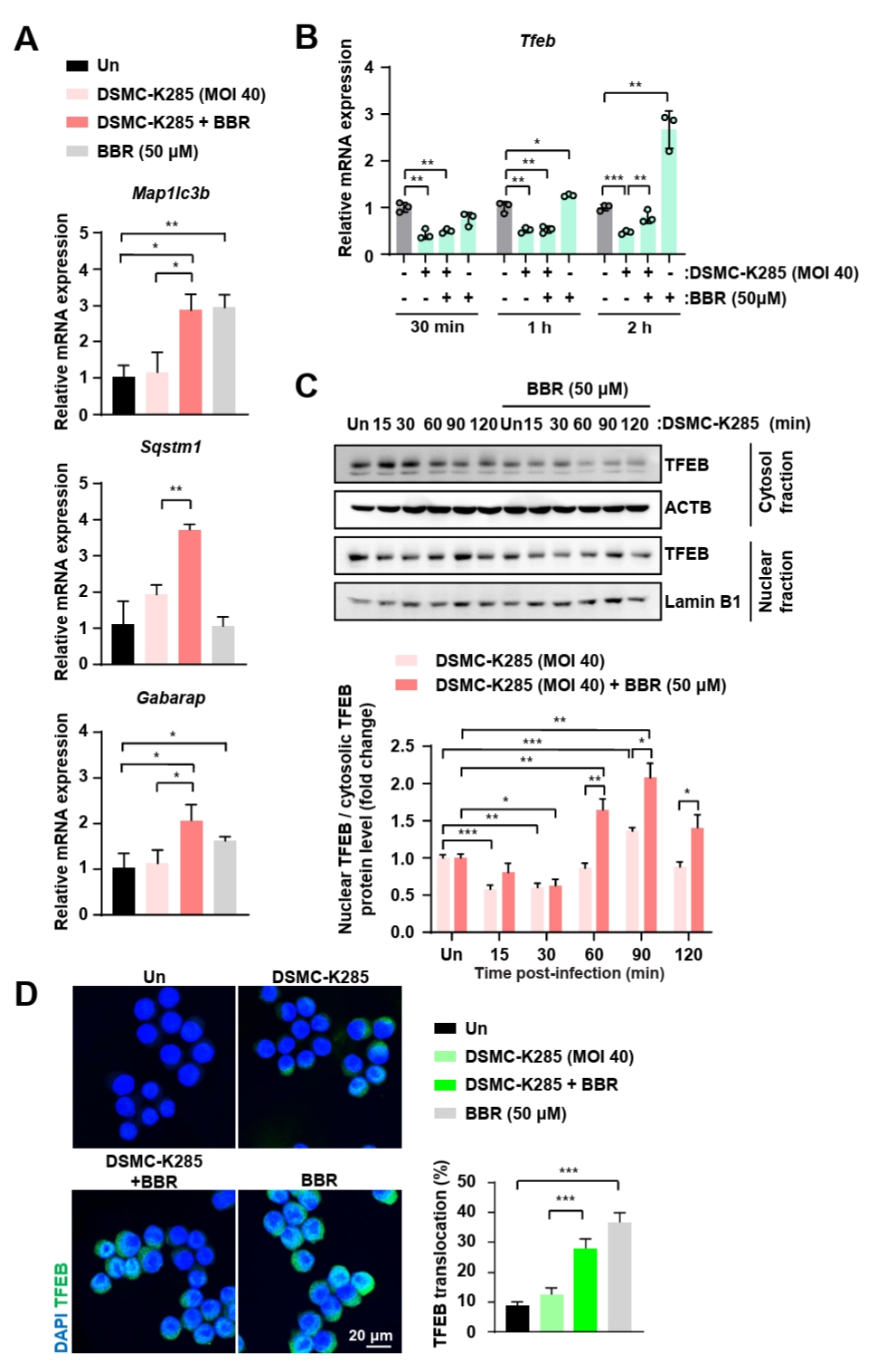
TFEB is a master regulator of autophagy and lysosomal biogenesis, and has been implicated in the regulation of lipophagy and lipolysis (Li et al., 2025b). Moreover, berberine modulates the activation of TFEB in macrophages (Zheng et al., 2021). As berberine decreased LD formation in hvKp-infected macrophages, we investigated whether TFEB signaling was involved in this process. To this end, we measured the mRNA levels of autophagy-related genes, including microtubule-associated protein 1 light chain 3 beta (LC3; encoded by Map1lc3b), sequestosome 1 (p62; encoded by Sqstm1), and GABA type A receptor-associated protein (Gabarap). Berberine co-treatment significantly increased the mRNA expression of these autophagy-related genes in hvKp-infected RAW 264.7 cells (Fig. 4A). We next evaluated Tfeb mRNA expression following hvKp infection and found that Tfeb expression levels were markedly decreased 2 h post-infection and were restored by berberine treatment (Fig. 4B). To determine whether berberine promotes TFEB nuclear translocation, we performed nuclear and cytoplasmic fractionation to assess the subcellular distribution of TFEB. Berberine treatment enhanced TFEB nuclear accumulation in hvKp-infected murine macrophages, as shown by fractionation and imaging analyses (Fig. 4C–4D). Collectively, these data demonstrate that berberine promotes TFEB activation, as evidenced by its enhanced nuclear translocation, in hvKp-infected murine macrophages.
Berberine does not restore hvKp-induced suppression of autophagy and autophagic flux in macrophages
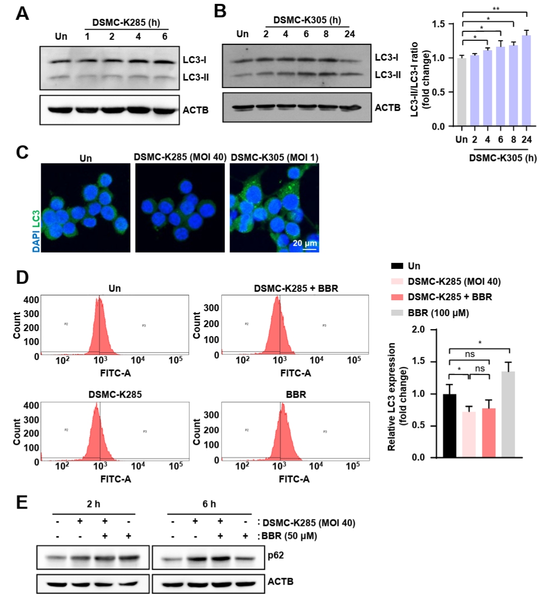
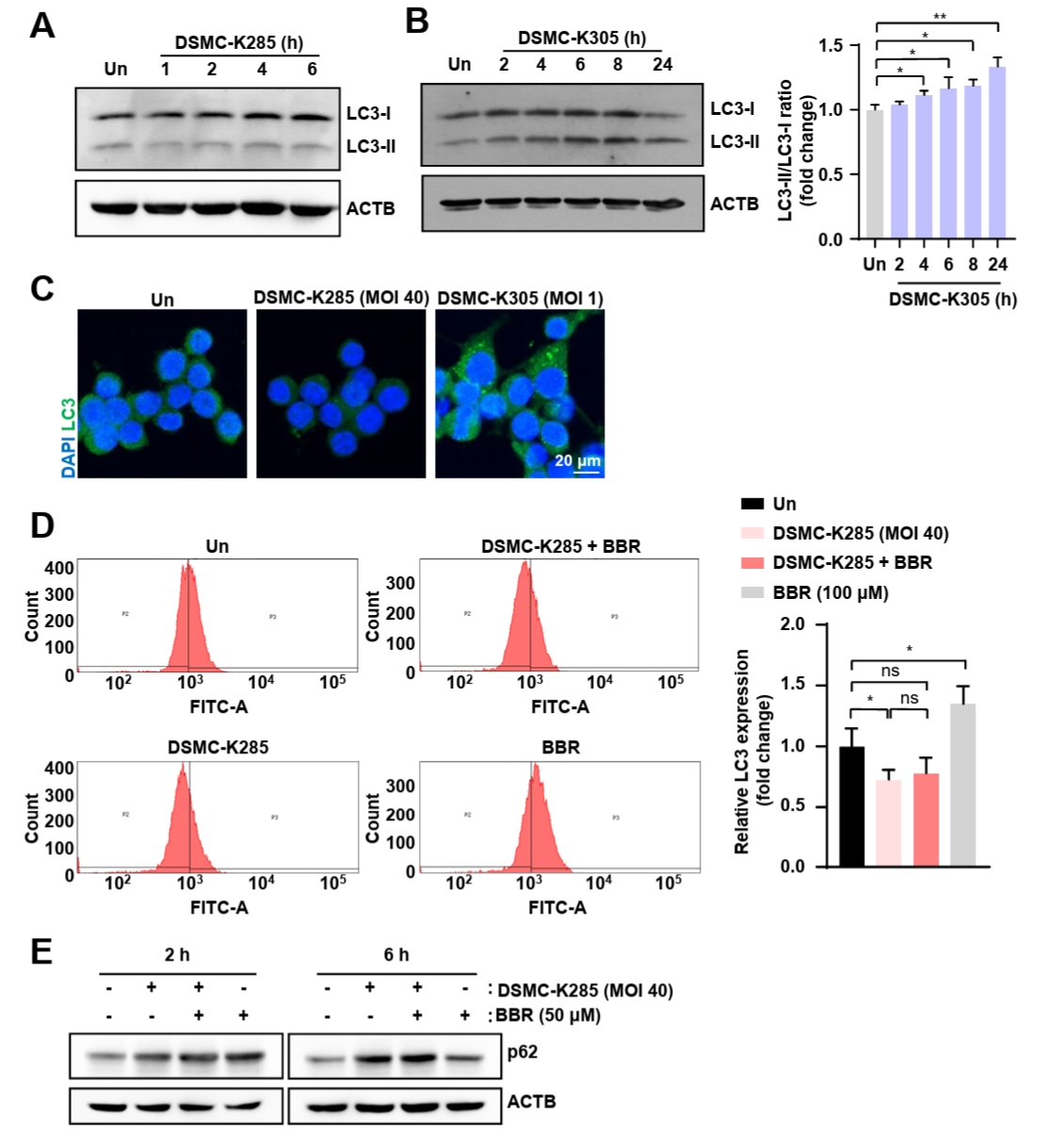
Berberine has been shown to reduce LD in various cell types by modulating lipid metabolic pathways (Ren et al., 2020; Wang et al., 2025). Here, we investigated whether berberine removes LD through autophagy activation. We examined the protein levels of LC3, a key autophagy-related protein, and found that LC3 levels were not significantly increased in hvKp-infected murine macrophages compared with those in uninfected controls, suggesting that autophagy was not activated during infection (Fig. 5A). To determine whether this lack of autophagy induction was specific to hvKp, we compared LC3 expression between hvKp- and cKp-infected macrophages. In contrast to hvKp infection, LC3 expression in cKp-infected macrophages gradually increased over time (Fig. 5B). Confocal microscopy confirmed that LC3 expression was significantly higher in cKp-infected macrophages than in hvKp-infected macrophages (Fig. 5C). Berberine treatment failed to induce LC3 expression in hvKp-infected murine macrophages (Fig. 5D). To further assess autophagic flux, we examined p62 levels and confirmed p62 protein levels did not decrease over time in either the hvKp-infected or berberine co-treated groups, indicating that autophagic flux remained impaired and was not functionally restored by berberine (Fig. 5E). Collectively, these results indicate that hvKp, unlike cKp, suppresses autophagy induction and impairs autophagic flux in macrophages, and that berberine co-treatment is insufficient to restore functional autophagy under these conditions.
Berberine enhances lipolysis in hvKp-infected murine macrophages
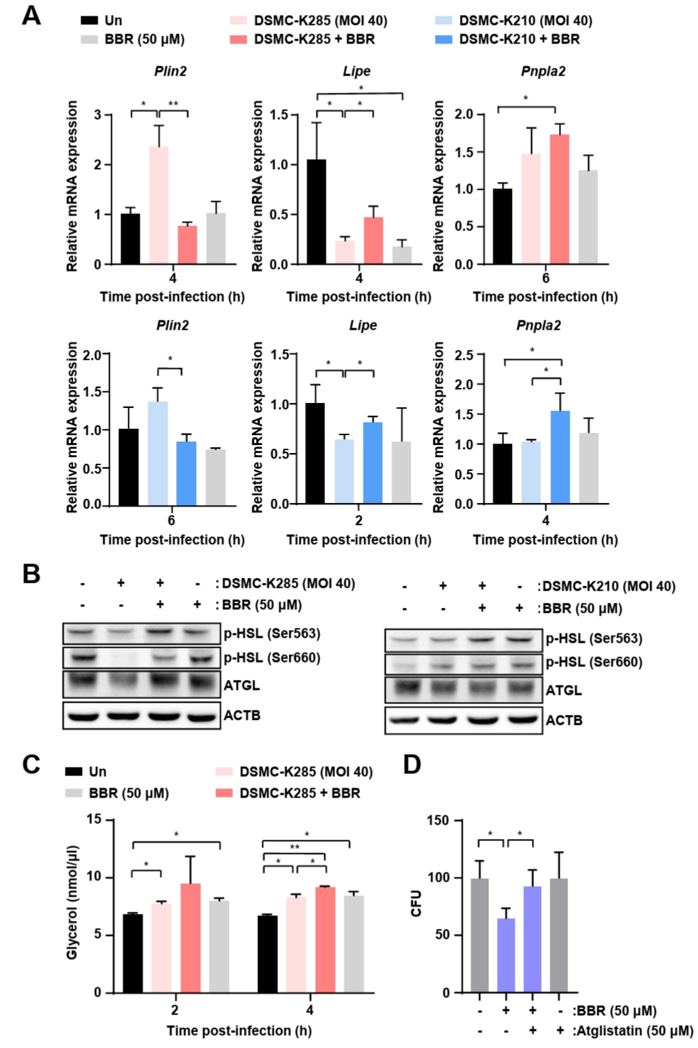
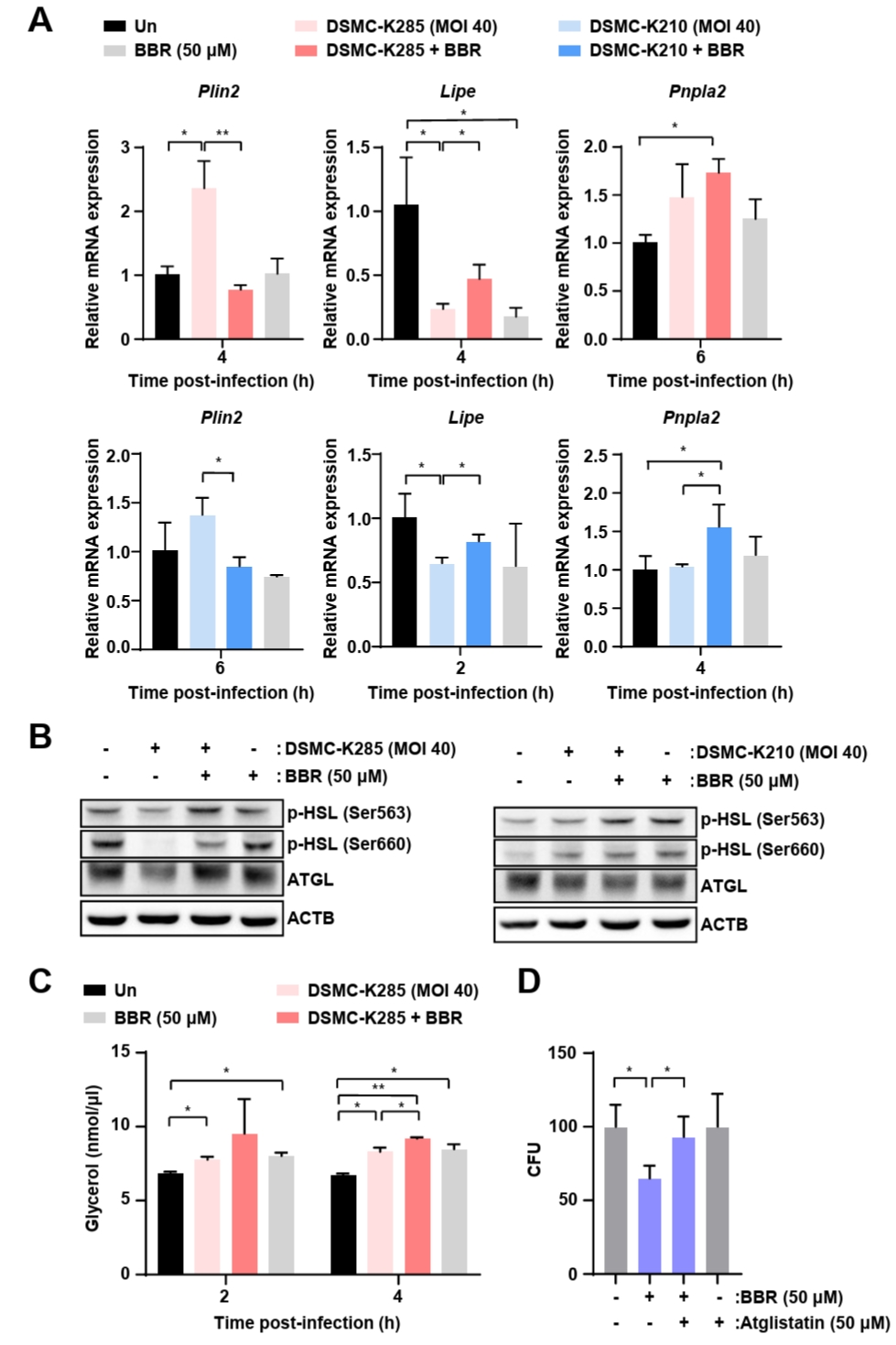
To investigate the mechanism by which berberine regulates lipid metabolism during hvKp infection, we focused on the lipolysis pathway and measured the mRNA levels of perilipin 2 (PLIN2; encoded by Plin2), a LD coat protein that restricts lipase access to LD, along with two key lipolytic enzymes: hormone-sensitive lipase (HSL; encoded by Lipe) and adipose triglyceride lipase (ATGL; encoded by Pnpla2). Compared with hvKp-infected cells, berberine-treated cells showed a significant increase in the mRNA expression of Pnpla2 and Lipe. In contrast, the expression of Plin2 was markedly decreased (Fig. 6A). Berberine treatment restored ATGL protein expression, which was reduced by hvKp infection (Fig. 6B). Berberine treatment also increased the phosphorylation of HSL at Ser563 and Ser660, indicating activation of the lipolytic enzyme cascade (Fig. 6B). Berberine treatment increased the level of free glycerol compared with hvKp infection alone, indicating enhanced TG breakdown through lipolysis (Fig. 6C). To determine whether ATGL-mediated lipolysis is required for the protective effect of berberine, we co-treated hvKp-infected macrophages with atglistatin, a selective ATGL inhibitor. Atglistatin treatment abolished the berberine-induced reduction in intracellular bacterial survival, indicating that ATGL-dependent lipolysis is required for berberine-mediated suppression of hvKp survival (Fig. 6D). Collectively, these results indicate that berberine promotes lipolysis, rather than lipophagy, as the primary mechanism through which it modulates lipid metabolism and suppresses LD accumulation during hvKp infection.
Discussion
Berberine is a plant-derived isoquinoline alkaloid extracted from Berberis vulgaris (barberry), Coptis chinensis (Chinese goldthread), and Hydrastis canadensis (goldenseal) (Cicero and Baggioni, 2016). It exhibits diverse pharmacological activities, including anti-inflammatory, antimicrobial, and metabolic regulatory effects (Gasmi et al., 2024). In a mouse model of Salmonella enterica serovar Typhimurium infection, berberine alleviated enteritis and protected against endotoxic shock (Li et al., 2022). Berberine exhibits significant antimicrobial activity against methicillin-resistant Staphylococcus aureus isolated from patients with bloodstream infections (Xia et al., 2022). Although its protective effects have been demonstrated in several infectious disease models, its role in host metabolic regulation during hvKp infection has not yet been defined. In this study, we demonstrated that berberine significantly reduced intracellular hvKp survival in macrophages and modulated infection-associated lipid metabolic remodeling. These findings provide mechanistic insights into how host-directed metabolic intervention may influence hvKp pathogenesis.
Accumulating evidence indicates that metabolic reprogramming is a critical determinant of host–pathogen interactions (Gleeson and Sheedy, 2016; O'Neill and Pearce, 2016). LDs, once considered inert lipid storage organelles, are now recognized as dynamic regulators of immune responses and intracellular pathogen survival. For instance, in Mycobacterium tuberculosis infection, restricting fatty acid uptake by macrophages limits bacterial growth (Simwela et al., 2025). Similarly, during hvKp infection, LD formation functions as an energy reservoir and plays a critical role in supporting the survival of intracellular bacteria (Jung et al., 2024). However, the mechanisms by which host lipid metabolism can be therapeutically targeted to restrict hvKp survival remain poorly understood. In the present study, we demonstrated that berberine markedly suppressed LD accumulation in hvKp-infected macrophages, thereby limiting intracellular bacterial survival. Mechanistically, our findings suggest that berberine restricts hvKp replication by altering host lipid metabolism and reducing lipid availability in infected macrophages.
LD turnover in macrophages can be regulated through two major pathways: lipophagy and lipolysis. Lipophagy is the selective autophagic degradation of LD via lysosomal pathways and plays a crucial role in maintaining cellular lipid homeostasis and energy balance (Khawar et al., 2019). Lipolysis is an enzymatic breakdown process in which TGs are hydrolyzed into free fatty acids and glycerol (Lass et al., 2011). Previous studies have shown that Kp infection impairs autophagic responses in macrophages (Wang et al., 2023), and berberine has been reported to regulate sirtuin 3-mediated lipophagy (Chen et al., 2025). Based on these observations, we initially hypothesized that berberine reduces LD accumulation by restoring lipophagy during hvKp infection. Consistent with this possibility, berberine treatment increased the expression of autophagy-related genes. TFEB, a key transcription factor that promotes autophagy (Zheng et al., 2021), was suppressed during hvKp infection but was restored following berberine treatment. Despite these transcriptional changes, autophagic flux remained largely unchanged, indicating that lipophagy was not functionally restored under these conditions. This unexpected finding suggests that LD reduction during hvKp infection occurs independently of lipophagy. Instead, our results suggest an alternative mechanism involving lipolysis. Supporting this interpretation, previous studies have shown that TFEB can regulate lipolytic pathways in addition to lipophagy (Li et al., 2021) and that berberine induces lipolysis in adipocytes by activating the AMP-activated protein kinase (AMPK) pathway (Yang et al., 2020). Taken together, our findings reveal that berberine limits intracellular hvKp survival by promoting lipolysis rather than lipophagy, thereby reducing the host lipid availability required for bacterial persistence. These results highlight the previously underappreciated role of lipolysis in controlling infection-induced LD accumulation, and suggest that targeting host lipid metabolism may represent a potential host-directed therapeutic strategy against hvKp infection.
Additionally, inflammatory responses play a crucial role in host defense by promoting pathogen clearance (Cronkite and Strutt, 2018). Excessive inflammation can lead to tissue damage (Paludan et al., 2021). HvKp infection triggers robust inflammatory responses in macrophages by activating the spleen tyrosine kinase-dependent immune-responsive gene 1-itaconate pathway (Mina et al., 2024) and inducing mitochondrial damage that promotes NOD-, LRR-, and pyrin domain-containing protein 3 inflammasome activation (Li et al., 2025a), highlighting the importance of maintaining homeostasis. Previous studies have shown that berberine exerts anti-inflammatory effects on macrophages (Liu et al., 2018; Xiong et al., 2023). Consistent with these reports, we observed that berberine effectively attenuated hvKp-induced inflammatory responses. Berberine is known to modulate MAPK signaling in macrophages under inflammatory conditions by activating AMPK (Jeong et al., 2009). Our findings indicate that berberine suppresses the activation of the JNK and ERK pathways induced by hvKp infection. This inhibition likely reduces pro-inflammatory cytokine production, limits excessive immune-mediated tissue damage, and preserves host defense.
Berberine has been reported to reduce intracellular lipid accumulation and oxidative stress through modulation of Lrrc58a in zebrafish hepatocytes (Wang et al., 2026), and a similar lipid-regulatory mechanism may contribute to the restoration of macrophage bactericidal function observed in our in vitro model. In the zebrafish system, berberine has likewise been shown to suppress macrophage and neutrophil recruitment and to downregulate TNF-α, IL-1β, and IL-6 expression (Zhang et al., 2020). Given the evolutionary conservation of NF-κB and related inflammatory signaling pathways, these findings collectively suggest that berberine may exert its protective effects through a conserved innate immune axis across both experimental systems.
Several limitations should be considered. First, this study was conducted in RAW 264.7 murine macrophages, which may not fully represent primary or tissue-resident macrophage responses. Second, although hvKp infection was associated with reduced Tfeb expression and impaired autophagy-related responses, the upstream signaling mechanisms remain to be clarified. Third, additional mechanistic in vivo validation is required to determine whether berberine-mediated lipolysis restricts bacterial survival during systemic infection. Despite these limitations, our results highlight that lipid metabolic regulation is a key determinant of hvKp intracellular survival. By promoting lipolysis and attenuating MAPK-driven inflammation, berberine disrupts the metabolic conditions favorable for bacterial persistence while moderating excessive host responses. These findings support further investigation of berberine as a potential host-directed therapeutic strategy targeting the metabolic pathways involved in hvKp pathogenesis.
Acknowledgments
The authors are thankful for all the helpful discussions and technical assistance. We also appreciate the contributions of the Core Facility Center at the School of Medicine, Keimyung University for instrument access and technical guidance.
This work was supported by the National Research Foundation of Korea (NRF) through the Sejong Science Fellowship (NRF-2022R1C1C2010228); by the Korea Basic Science Institute under the R&D Program (Project No. C523311) funded by the Ministry of Science and ICT.
Conflict of Interest
The authors have no conflict of interest to report.
Ethical Statements
All ZF experiments were conducted in accordance with protocols approved by the Animal Research Ethics Committee of Gyeongsang National University (approval GNU-190325-E0014).
Berberine treatment shows no effect on hvKp growth. HvKp (DSMC-K285 or DSMCK210, MOI 40) were incubated with berberine (50 or 100 μM) at various time points in culture medium. All data expressed as the Mean ± SD from three independent experiments (n = 3). Statistical significance was determined using Student’s t-test. *P < 0.05, **P < 0.005, and ***P < 0.001 according to analysis of variance. ns, not significant; BBR, berberine.
Berberine inhibits intracellular hvKp survival. (A) Chemical structure of berberine. (B) RAW 264.7 cells were treated with berberine (10, 25, or 50 μM) for 6 h, and cell viability was assessed using the MTS assay. (C) RAW 264.7 cells were infected with hvKp (DSMC-K210 or DSMC-K285, MOI 40) and then treated with berberine (50 μM) for 6 h. Cell viability was subsequently evaluated using the MTS assay. (D) RAW 264.7 cells were infected with hvKp (DSMC-K210 or DSMC-K285, MOI 40) and treated with berberine (50 μM) for the indicated times. Intracellular bacterial burden was measured using the CFU assay. (E) Representative images of GFP-expressing hvKp infection in ZF larvae. Fluorescence intensity was reduced in larvae treated with berberine or AMK compared with the untreated infected group. (F) Bacterial burden (log10 CFU/larva) in hvKp-infected ZF larvae treated with berberine or AMK. (G) Kaplan–Meier survival curves of hvKp-infected ZF larvae treated with berberine or AMK. Zebrafish larvae were infected at 3 days post-fertilization (dpf), followed by antibiotic treatment, and survival was monitored for 7 days post-injection (dpi). Survival was prolonged in the berberine- and AMK-treated groups compared with the untreated infected group. Data in panels B–D are expressed as the Mean ± SD from three independent experiments (n = 3). Statistical significance was determined using Student’s t-test. *P < 0.05, **P < 0.005, ***P < 0.001. Data in panel F are presented as the Mean ± SEM (n = 10 larvae/group). Statistical significance was determined by one-way ANOVA followed by Dunnett’s multiple comparisons test, with p-values calculated relative to the untreated infected control group (Un), where ***P < 0.001. Un, untreated (control group); ns, not significant; AMK, amikacin; BBR, berberine.
Fig. 2.
Berberine reduces inflammation in hvKp-infected macrophages. (A) RAW 264.7 cells infected with hvKp (DSMC-K210 or DSMC-K285, MOI 40) were treated with berberine (50 μM) for 4 h, and pro-inflammatory cytokine mRNA levels were measured by RT-qPCR. (B) RAW 264.7 cells infected with hvKp (DSMC-K285, MOI 40) were treated with berberine (50 or 100 μM), and protein levels of TNF-α and IL-6 in the culture supernatants were measured by ELISA. (C) RAW 264.7 cells were infected with hvKp (DSMC-K285, MOI 40), treated with berberine (50 μM), and harvested at the indicated time points for Western blot analysis of ERK and JNK phosphorylation. ACTB was used as the loading control. All quantitative data are expressed as the Mean ± SD from three independent experiments (n = 3). Statistical significance was determined using Student’s t-test. *P < 0.05, **P < 0.005, ***P < 0.001. Un, untreated (control group); ns, not significant; BBR, berberine.
Fig. 3.
Berberine reduces hvKp-induced LD accumulation in macrophages. (A) RAW 264.7 cells were infected with hvKp (DSMC-K285, MOI 40) and treated with berberine (50 μM) for 6 h, and mTOR signaling was assessed by Western blot. ACTB was used as the loading control. (B) RAW 264.7 cells were infected with hvKp (DSMC-K285, MOI 40) and treated with berberine (50 μM) for 4 h. The expression levels of lipogenesis-related genes (Fasn, Scd1, and Acaca) were analyzed using RT-qPCR. (C) LD formation in RAW 264.7 cells under hvKp infection and berberine treatment was visualized by confocal microscopy. Images are representative of three independent experiments. Scale bar = 20 μm. (D) RAW 264.7 cells were infected with hvKp (DSMC-K210) and treated with berberine (50 μM), and PLIN1 expression was assessed by Western blotting. ACTB was used as the loading control. (E) Intracellular TG levels were quantified in RAW 264.7 cells following hvKp (DSMC-K285) infection and berberine treatment (100 μM). (F) LD in RAW 264.7 cells under hvKp infection and berberine treatment were visualized by TEM and quantified as shown in the panel. Yellow arrows indicate LD. Scale bars = 2 and 5 μm. All quantitative data are expressed as the Mean ± SD from three independent experiments (n = 3). Data in panel F are expressed as the Mean ± SD (n = 30). Statistical significance was determined using Student’s t-test. *P < 0.05, **P < 0.005, ***P < 0.001. Un, uninfected (control group); LD, lipid droplet; TG, triglyceride; N, nucleus; BBR, berberine.
Fig. 4.
Berberine increases TFEB expression and nuclear accumulation in hvKp-infected macrophages. (A) RAW 264.7 cells were infected with hvKp (DSMC-K285) and treated with berberine (50 μM) for 4 h, and autophagy-related gene expression was measured by RT-qPCR. (B) HvKp-infected RAW 264.7 cells were treated with berberine (50 μM), and Tfeb mRNA levels were analyzed by RT-qPCR at the indicated time points. (C) TFEB protein levels in nuclear and cytosolic fractions were assessed by Western blotting after berberine treatment at the indicated time points. ACTB and lamin B1 were used as cytosolic and nuclear fraction controls, respectively. (D) Confocal microscopy was performed to visualize the nuclear translocation of TFEB following berberine treatment. Images are representative of three independent experiments. Scale bar = 20 μm. Data are expressed as the Mean ± SD from three independent experiments (n = 3). Statistical significance was determined using Student’s t-test. *P < 0.05, **P < 0.005, ***P < 0.001. Un, uninfected (control group); BBR, berberine.
Fig. 5.
Berberine co-treatment fails to restore autophagy induction and autophagic flux impaired by hvKp infection in macrophages. (A–C) RAW 264.7 cells were infected with hvKp (DSMC-K285, MOI 40) or cKp (DSMC-K305, MOI 1). (A and B) LC3 protein expression was analyzed by Western blotting using ACTB as the loading control. (C) Representative confocal images showing LC3 expression. Scale bar = 20 μm. (D) RAW 264.7 cells infected with hvKp (DSMC-K285) and treated with berberine (100 μM) were analyzed for LC3 fluorescence by flow cytometry. (E) RAW 264.7 cells infected with hvKp (DSMC-K285, MOI 40) and treated with berberine (50 μM). p62 expression was analyzed by Western blot at 2 and 6 h post-infection. Images are representative of three independent experiments. Data are expressed as the Mean ± SD from three independent experiments (n = 3). Statistical significance was determined using Student’s t-test. *P < 0.05, **P < 0.005, ***P < 0.001. Un, uninfected (control group); ns, not significant; BBR, berberine.
Fig. 6.
Berberine promotes lipolysis-associated responses in hvKp-infected macrophages. (A) RAW 264.7 cells were infected with hvKp (DSMC-K210 or DSMC-K285, MOI 40) and treated with berberine (50 μM), and the mRNA levels of lipolysis-related genes were measured by RT-qPCR. (B) RAW 264.7 cells were infected with hvKp (DSMC-K210 or DSMC-K285, MOI 40) and treated with berberine (50 μM), and the protein levels of lipolysis-related proteins were measured by Western blot. (C) RAW 264.7 cells were infected with hvKp (DSMC-K285, MOI 40) and treated with berberine (50 μM), and free glycerol levels in the culture supernatant were quantified. (D) RAW 264.7 cells were infected with hvKp (DSMC-K285, MOI 40) and treated with berberine (50 μM) in the presence or absence of the ATGL inhibitor atglistatin (50 μM), and intracellular bacterial burden was measured after 24 h by CFU assay. All data are expressed as the Mean ± SD from three independent experiments (n = 3). Statistical significance was determined using Student’s t-test. *P < 0.05, **P < 0.005, ***P < 0.001. Un, untreated (control group); BBR, berberine.
References
Abbas R, Chakkour M, Zein El Dine H, Obaseki EF, Obeid ST, et al. 2024. General overview of Klebsiella pneumoniae: Epidemiology and the role of siderophores in its pathogenicity. Biology (Basel). 13: 78.ArticlePubMedPMC
Bolsoni-Lopes A, Alonso-Vale MI. 2015. Lipolysis and lipases in white adipose tissue - An update. Arch Endocrinol Metab. 59: 335–342. ArticlePubMed
Cai Y, Yang Q, Yu Y, Yang F, Bai R, et al. 2023. Efficacy and underlying mechanisms of berberine against lipid metabolic diseases: A review. Front Pharmacol. 14: 1283784.ArticlePubMedPMC
Chen W, Jin T, Xie Y, Zhong C, Gao H, et al. 2025. Berberine partially ameliorates cardiolipotoxicity in diabetic cardiomyopathy by modulating SIRT3-mediated lipophagy to remodel lipid droplets homeostasis. Br J Pharmacol. 182: 5038–5056. ArticlePubMed
Cicero AF, Baggioni A. 2016. Berberine and its role in chronic disease. Adv Exp Med Biol. 928: 27–45. ArticlePubMed
Corbo JH, Chung J. 2024. Mechanisms of lipid droplet degradation. Curr Opin Cell Biol. 90: 102402.ArticlePubMed
Cronkite DA, Strutt TM. 2018. The regulation of inflammation by innate and adaptive lymphocytes. J Immunol Res. 2018: 1467538.ArticlePubMedPMCPDF
Düvel K, Yecies JL, Menon S, Raman P, Lipovsky AI, et al. 2010. Activation of a metabolic gene regulatory network downstream of mTOR complex 1. Mol Cell. 39: 171–183. ArticlePubMedPMC
Gasmi A, Asghar F, Zafar S, Oliinyk P, Khavrona O, et al. 2024. Berberine: Pharmacological features in health, disease and aging. Curr Med Chem. 31: 1214–1234. ArticlePubMedLink
Gleeson LE, Sheedy FJ. 2016. Metabolic reprogramming & inflammation: Fuelling the host response to pathogens. Semin Immunol. 28: 450–468. ArticlePubMed
Han J, Wang Y. 2018. mTORC1 signaling in hepatic lipid metabolism. Protein Cell. 9: 145–151. ArticlePubMedPMCPDF
Jeon SM, Kim YJ, Nguyen TQ, Cui J, Thi Bich Hanh B, et al. 2022. Ohmyungsamycin promotes M1-like inflammatory responses to enhance host defence against Mycobacteroides abscessus infections. Virulence. 13: 1966–1984. ArticlePubMedPMC
Jeong HW, Hsu KC, Lee JW, Ham M, Huh JY, et al. 2009. Berberine suppresses proinflammatory responses through AMPK activation in macrophages. Am J Physiol Endocrinol Metab. 296: E955–E964. ArticlePubMed
Jung HJ, Kim HA, Hyun M, Lee JY, Kim YJ, et al. 2024. Inhibiting lipid droplet biogenesis enhances host protection against hypervirulent Klebsiella pneumoniae infections. Med Microbiol Immunol. 213: 26.ArticlePubMedPMCPDF
Khawar MB, Gao H, Li W. 2019. Autophagy and lipid metabolism. Adv Exp Med Biol. 1206: 359–374. ArticlePubMed
Kounakis K, Chaniotakis M, Markaki M, Tavernarakis N. 2019. Emerging roles of lipophagy in health and disease. Front Cell Dev Biol. 7: 185.Article
Lass A, Zimmermann R, Oberer M, Zechner R. 2011. Lipolysis - A highly regulated multi-enzyme complex mediates the catabolism of cellular fat stores. Prog Lipid Res. 50: 14–27. ArticlePubMedPMC
Leopold Wager CM, Arnett E, Schlesinger LS. 2019. Macrophage nuclear receptors: Emerging key players in infectious diseases. PLoS Pathog. 15: e1007585. ArticlePubMedPMC
Li Y, Li X, Wu W, Liu P, Liu J, et al. 2025a. Insights into the roles of macrophages in Klebsiella pneumoniae infections: A comprehensive review. Cell Mol Biol Lett. 30: 34.ArticlePubMedPMCPDF
Li L, Lin J, Huang C, Liu J, Yuan Y, et al. 2025b. The TFEB activator clomiphene citrate ameliorates lipid metabolic syndrome pathology by activating lipophagy and lipolysis. Biochem Pharmacol. 232: 116694.Article
Li M, Wang Z, Wang P, Li H, Yang L. 2021. TFEB: A emerging regulator in lipid homeostasis for atherosclerosis. Front Physiol. 12: 639920.ArticlePubMedPMC
Li CG, Yan L, Jing YY, Xu LH, Liang YD, et al. 2017. Berberine augments ATP-induced inflammasome activation in macrophages by enhancing AMPK signaling. Oncotarget. 8: 95–109. ArticlePubMedPMC
Li M, Zhang H, Zhang Y, Fan J, Zhu J, et al. 2022. Berberine modulates macrophage activation by inducing glycolysis. J Immunol. 208: 2309–2318. ArticlePubMedPDF
Liu Y, Liu X, Hua W, Wei Q, Fang X, et al. 2018. Berberine inhibits macrophage M1 polarization via AKT1/SOCS1/NF-κB signaling pathway to protect against DSS-induced colitis. Int Immunopharmacol. 57: 121–131. ArticlePubMed
Liu X, Xu Q, Yang X, Heng H, Yang C, et al. 2025. Capsular polysaccharide enables Klebsiella pneumoniae to evade phagocytosis by blocking host-bacteria interactions. mBio. 16: e0383824. ArticlePubMedPMCLink
March C, Moranta D, Regueiro V, Llobet E, Tomás A, et al. 2011. Klebsiella pneumoniae outer membrane protein A is required to prevent the activation of airway epithelial cells. J Biol Chem. 286: 9956–9967. ArticlePubMedPMC
Marcoleta AE, Varas MA, Ortiz-Severín J, Vásquez L, Berríos-Pastén C, et al. 2018. Evaluating different virulence traits of Klebsiella pneumoniae using Dictyostelium discoideum and zebrafish larvae as host models. Front Cell Infect Microbiol. 8: 30.ArticlePubMedPMC
Marr CM, Russo TA. 2019. Hypervirulent Klebsiella pneumoniae: A new public health threat. Expert Rev Anti Infect Ther. 17: 71–73. ArticlePubMed
Mashek DG. 2021. Hepatic lipid droplets: A balancing act between energy storage and metabolic dysfunction in NAFLD. Mol Metab. 50: 101115.ArticlePubMed
Mina SA, Zhu G, Fanian M, Chen S, Yang G. 2024. Exploring reduced macrophage cell toxicity of hypervirulent Klebsiella pneumoniae compared to classical Klebsiella pneumoniae. Microbiol Res. 278: 127515.ArticlePubMed
Mohd Asri NA, Ahmad S, Mohamud R, Mohd Hanafi N, Mohd Zaidi NF, et al. 2021. Global prevalence of nosocomial multidrug-resistant Klebsiella pneumoniae: A systematic review and meta-analysis. Antibiotics (Basel). 10: 1508.ArticlePubMedPMC
Neag MA, Mocan A, Echeverría J, Pop RM, Bocsan CI, et al. 2018. Berberine: Botanical occurrence, traditional uses, extraction methods, and relevance in cardiovascular, metabolic, hepatic, and renal disorders. Front Pharmacol. 9: 557.ArticlePubMedPMC
Nguyen TQ, Heo BE, Hanh BTB, Jeon S, Park Y, et al. 2023. DS86760016, a leucyl-tRNA synthetase inhibitor, is active against Mycobacterium abscessus. Antimicrob Agents Chemother. 67: e0156722. ArticlePubMedPMCLink
Olzmann JA, Carvalho P. 2019. Dynamics and functions of lipid droplets. Nat Rev Mol Cell Biol. 20: 137–155. ArticlePubMedPMCPDF
Ren G, Guo JH, Qian YZ, Kong WJ, Jiang JD. 2020. Berberine improves glucose and lipid metabolism in HepG2 cells through AMPKα1 activation. Front Pharmacol. 11: 647.ArticlePubMedPMC
Russo TA, Marr CM. 2019. Hypervirulent Klebsiella pneumoniae. Clin Microbiol Rev. 32: e00001-19.ArticlePubMedPMCLink
Simwela NV, Jaecklein E, Sassetti CM, Russell DG. 2025. Impaired fatty acid import or catabolism in macrophages restricts intracellular growth of Mycobacterium tuberculosis. eLife. 13: RP102980.ArticlePubMedPMCPDF
Singh R, Kaushik S, Wang Y, Xiang Y, Novak I, et al. 2009. Autophagy regulates lipid metabolism. Nature. 458: 1131–1135. ArticlePubMedPMCPDF
Takaki K, Davis JM, Winglee K, Ramakrishnan L. 2013. Evaluation of the pathogenesis and treatment of Mycobacterium marinum infection in zebrafish. Nat Protoc. 8: 1114–1124. ArticlePubMedPMCPDF
Tian X, Xie G, Xiao H, Ding F, Bao W, et al. 2019. CXCR4 knockdown prevents inflammatory cytokine expression in macrophages by suppressing activation of MAPK and NF-κB signaling pathways. Cell Biosci. 9: 55.ArticlePubMedPMCPDF
Vrieling F, Wilson L, Rensen PCN, Walzl G, Ottenhoff THM, et al. 2019. Oxidized low-density lipoprotein (oxLDL) supports Mycobacterium tuberculosis survival in macrophages by inducing lysosomal dysfunction. PLoS Pathog. 15: e1007724. ArticlePubMedPMC
Wang X, Bi C, Xin X, Zhang M, Fu H, et al. 2023. Pyroptosis, apoptosis, and autophagy are involved in infection induced by two clinical Klebsiella pneumoniae isolates with different virulence. Front Cell Infect Microbiol. 13: 1165609.ArticlePubMedPMC
Wang X, Kin Jacky HC, Fp B, Che Y, Sun Z, et al. 2026. Berberine regulates cellular lipid accumulation, oxidative stress, and cell proliferation by modulating Lrrc58a in sodium palmitate-induced zebrafish hepatocyte. AIMS Anim Sci. 2: 35–52. Article
Wang S, Liu R, Yu Q, Dong L, Bi Y, et al. 2019. Metabolic reprogramming of macrophages during infections and cancer. Cancer Lett. 452: 14–22. ArticlePubMed
Wang H, Wang Z, Wang D, Nie K, Wu W, et al. 2025. Berberine attenuates nonalcoholic hepatic steatosis by regulating lipid droplet-associated proteins: In vivo, in vitro and molecular evidence. J Cell Mol Med. 29: e70524.ArticlePubMedPMCLink
Wang G, Zhao G, Chao X, Xie L, Wang H. 2020. The characteristic of virulence, biofilm and antibiotic resistance of Klebsiella pneumoniae. Int J Environ Res Public Health. 17: 6278.ArticlePubMedPMC
Wu JH, Hong LC, Tsai YY, Chen HW, Chen WX, et al. 2006. Mitogen-activated protein kinase (MAPK) signalling pathways in HepG2 cells infected with a virulent strain of Klebsiella pneumoniae. Cell Microbiol. 8: 1467–1474. ArticlePubMed
Xia S, Ma L, Wang G, Yang J, Zhang M, et al. 2022. In vitro antimicrobial activity and the mechanism of berberine against methicillin-resistant Staphylococcus aureus isolated from bloodstream infection patients. Infect Drug Resist. 15: 1933–1944. ArticlePubMedPMCLink
Xiong K, Deng J, Yue T, Hu W, Zeng X, et al. 2023. Berberine promotes M2 macrophage polarisation through the IL-4-STAT6 signalling pathway in ulcerative colitis treatment. Heliyon. 9: e14176. ArticlePubMedPMC
Xu G, Wang J, Liu CH. 2017. Klebsiella pneumoniae NdpA suppresses ERK pathway-mediated host early inflammatory responses and is degraded through the ubiquitin-proteasome pathway. Protein Cell. 8: 144–148. ArticlePubMedPMCPDF
Xu Q, Yang X, Chan EWC, Chen S. 2021. The hypermucoviscosity of hypervirulent K. pneumoniae confers the ability to evade neutrophil-mediated phagocytosis. Virulence. 12: 2050–2059. ArticlePubMedPMCLink
Yang Y, Lu R, Gao F, Zhang J, Liu F. 2020. Berberine induces lipolysis in porcine adipocytes by activating the AMP-activated protein kinase pathway. Mol Med Rep. 21: 2603–2614. ArticlePubMed
Ye J, Lu L, Rui XH, Ren MD, Tu F, et al. 2025. Zebrafish as a model for investigating Klebsiella pneumoniae-driven lung injury and therapeutic targets. Exp Lung Res. 51: 11–22. ArticlePubMed
Zechner R, Madeo F, Kratky D. 2017. Cytosolic lipolysis and lipophagy: Two sides of the same coin. Nat Rev Mol Cell Biol. 18: 671–684. ArticlePubMedPDF
Zechner R, Zimmermann R, Eichmann TO, Kohlwein SD, Haemmerle G, et al. 2012. FAT SIGNALS - Lipases and lipolysis in lipid metabolism and signaling. Cell Metab. 15: 279–291. ArticlePubMedPMC
Zhang B, Wang L, Ji X, Zhang S, Sik A, et al. 2020. Anti-inflammation associated protective mechanism of berberine and its derivatives on attenuating pentylenetetrazole-induced seizures in zebrafish. J Neuroimmune Pharmacol. 15: 309–325. ArticlePubMedPDF
Zhang S, Xu P, Zhu Z, Zhou L, Li J, et al. 2023. Acetylation of p65Lys310 by p300 in macrophages mediates anti-inflammatory property of berberine. Redox Biol. 62: 102704.ArticlePubMedPMC
Zheng Y, Kou J, Wang P, Ye T, Wang Z, et al. 2021. Berberine-induced TFEB deacetylation by SIRT1 promotes autophagy in peritoneal macrophages. Aging (Albany NY). 13: 7096–7119. ArticlePubMedPMC
Berberine promotes host lipolysis to enhance antimicrobial defense against hypervirulent Klebsiella pneumoniae infection
Fig. 1. Berberine inhibits intracellular hvKp survival. (A) Chemical structure of berberine. (B) RAW 264.7 cells were treated with berberine (10, 25, or 50 μM) for 6 h, and cell viability was assessed using the MTS assay. (C) RAW 264.7 cells were infected with hvKp (DSMC-K210 or DSMC-K285, MOI 40) and then treated with berberine (50 μM) for 6 h. Cell viability was subsequently evaluated using the MTS assay. (D) RAW 264.7 cells were infected with hvKp (DSMC-K210 or DSMC-K285, MOI 40) and treated with berberine (50 μM) for the indicated times. Intracellular bacterial burden was measured using the CFU assay. (E) Representative images of GFP-expressing hvKp infection in ZF larvae. Fluorescence intensity was reduced in larvae treated with berberine or AMK compared with the untreated infected group. (F) Bacterial burden (log10 CFU/larva) in hvKp-infected ZF larvae treated with berberine or AMK. (G) Kaplan–Meier survival curves of hvKp-infected ZF larvae treated with berberine or AMK. Zebrafish larvae were infected at 3 days post-fertilization (dpf), followed by antibiotic treatment, and survival was monitored for 7 days post-injection (dpi). Survival was prolonged in the berberine- and AMK-treated groups compared with the untreated infected group. Data in panels B–D are expressed as the Mean ± SD from three independent experiments (n = 3). Statistical significance was determined using Student’s t-test. *P < 0.05, **P < 0.005, ***P < 0.001. Data in panel F are presented as the Mean ± SEM (n = 10 larvae/group). Statistical significance was determined by one-way ANOVA followed by Dunnett’s multiple comparisons test, with p-values calculated relative to the untreated infected control group (Un), where ***P < 0.001. Un, untreated (control group); ns, not significant; AMK, amikacin; BBR, berberine.
Fig. 2. Berberine reduces inflammation in hvKp-infected macrophages. (A) RAW 264.7 cells infected with hvKp (DSMC-K210 or DSMC-K285, MOI 40) were treated with berberine (50 μM) for 4 h, and pro-inflammatory cytokine mRNA levels were measured by RT-qPCR. (B) RAW 264.7 cells infected with hvKp (DSMC-K285, MOI 40) were treated with berberine (50 or 100 μM), and protein levels of TNF-α and IL-6 in the culture supernatants were measured by ELISA. (C) RAW 264.7 cells were infected with hvKp (DSMC-K285, MOI 40), treated with berberine (50 μM), and harvested at the indicated time points for Western blot analysis of ERK and JNK phosphorylation. ACTB was used as the loading control. All quantitative data are expressed as the Mean ± SD from three independent experiments (n = 3). Statistical significance was determined using Student’s t-test. *P < 0.05, **P < 0.005, ***P < 0.001. Un, untreated (control group); ns, not significant; BBR, berberine.
Fig. 3. Berberine reduces hvKp-induced LD accumulation in macrophages. (A) RAW 264.7 cells were infected with hvKp (DSMC-K285, MOI 40) and treated with berberine (50 μM) for 6 h, and mTOR signaling was assessed by Western blot. ACTB was used as the loading control. (B) RAW 264.7 cells were infected with hvKp (DSMC-K285, MOI 40) and treated with berberine (50 μM) for 4 h. The expression levels of lipogenesis-related genes (Fasn, Scd1, and Acaca) were analyzed using RT-qPCR. (C) LD formation in RAW 264.7 cells under hvKp infection and berberine treatment was visualized by confocal microscopy. Images are representative of three independent experiments. Scale bar = 20 μm. (D) RAW 264.7 cells were infected with hvKp (DSMC-K210) and treated with berberine (50 μM), and PLIN1 expression was assessed by Western blotting. ACTB was used as the loading control. (E) Intracellular TG levels were quantified in RAW 264.7 cells following hvKp (DSMC-K285) infection and berberine treatment (100 μM). (F) LD in RAW 264.7 cells under hvKp infection and berberine treatment were visualized by TEM and quantified as shown in the panel. Yellow arrows indicate LD. Scale bars = 2 and 5 μm. All quantitative data are expressed as the Mean ± SD from three independent experiments (n = 3). Data in panel F are expressed as the Mean ± SD (n = 30). Statistical significance was determined using Student’s t-test. *P < 0.05, **P < 0.005, ***P < 0.001. Un, uninfected (control group); LD, lipid droplet; TG, triglyceride; N, nucleus; BBR, berberine.
Fig. 4. Berberine increases TFEB expression and nuclear accumulation in hvKp-infected macrophages. (A) RAW 264.7 cells were infected with hvKp (DSMC-K285) and treated with berberine (50 μM) for 4 h, and autophagy-related gene expression was measured by RT-qPCR. (B) HvKp-infected RAW 264.7 cells were treated with berberine (50 μM), and Tfeb mRNA levels were analyzed by RT-qPCR at the indicated time points. (C) TFEB protein levels in nuclear and cytosolic fractions were assessed by Western blotting after berberine treatment at the indicated time points. ACTB and lamin B1 were used as cytosolic and nuclear fraction controls, respectively. (D) Confocal microscopy was performed to visualize the nuclear translocation of TFEB following berberine treatment. Images are representative of three independent experiments. Scale bar = 20 μm. Data are expressed as the Mean ± SD from three independent experiments (n = 3). Statistical significance was determined using Student’s t-test. *P < 0.05, **P < 0.005, ***P < 0.001. Un, uninfected (control group); BBR, berberine.
Fig. 5. Berberine co-treatment fails to restore autophagy induction and autophagic flux impaired by hvKp infection in macrophages. (A–C) RAW 264.7 cells were infected with hvKp (DSMC-K285, MOI 40) or cKp (DSMC-K305, MOI 1). (A and B) LC3 protein expression was analyzed by Western blotting using ACTB as the loading control. (C) Representative confocal images showing LC3 expression. Scale bar = 20 μm. (D) RAW 264.7 cells infected with hvKp (DSMC-K285) and treated with berberine (100 μM) were analyzed for LC3 fluorescence by flow cytometry. (E) RAW 264.7 cells infected with hvKp (DSMC-K285, MOI 40) and treated with berberine (50 μM). p62 expression was analyzed by Western blot at 2 and 6 h post-infection. Images are representative of three independent experiments. Data are expressed as the Mean ± SD from three independent experiments (n = 3). Statistical significance was determined using Student’s t-test. *P < 0.05, **P < 0.005, ***P < 0.001. Un, uninfected (control group); ns, not significant; BBR, berberine.
Fig. 6. Berberine promotes lipolysis-associated responses in hvKp-infected macrophages. (A) RAW 264.7 cells were infected with hvKp (DSMC-K210 or DSMC-K285, MOI 40) and treated with berberine (50 μM), and the mRNA levels of lipolysis-related genes were measured by RT-qPCR. (B) RAW 264.7 cells were infected with hvKp (DSMC-K210 or DSMC-K285, MOI 40) and treated with berberine (50 μM), and the protein levels of lipolysis-related proteins were measured by Western blot. (C) RAW 264.7 cells were infected with hvKp (DSMC-K285, MOI 40) and treated with berberine (50 μM), and free glycerol levels in the culture supernatant were quantified. (D) RAW 264.7 cells were infected with hvKp (DSMC-K285, MOI 40) and treated with berberine (50 μM) in the presence or absence of the ATGL inhibitor atglistatin (50 μM), and intracellular bacterial burden was measured after 24 h by CFU assay. All data are expressed as the Mean ± SD from three independent experiments (n = 3). Statistical significance was determined using Student’s t-test. *P < 0.05, **P < 0.005, ***P < 0.001. Un, untreated (control group); BBR, berberine.
Fig. 1.
Fig. 2.
Fig. 3.
Fig. 4.
Fig. 5.
Fig. 6.
Berberine promotes host lipolysis to enhance antimicrobial defense against hypervirulent Klebsiella pneumoniae infection