- About
- Browse Articles
-
Special Issues
- Pioneering strategies for overcoming bacterial drug resistance (2026)
- Advancing microbial engineering through synthetic biology (2025)
- Host-associated microbiome (2024)
- Bacterial regulatory mechanisms for the control of complex cellular mechanisms (2023)
- Two years into COVID-19 pandemic: Where are we? (2022)
- Collections
- For Contributors
- Policies
- E-Submission

- About
- Browse Articles
-
Special Issues
- Pioneering strategies for overcoming bacterial drug resistance (2026)
- Advancing microbial engineering through synthetic biology (2025)
- Host-associated microbiome (2024)
- Bacterial regulatory mechanisms for the control of complex cellular mechanisms (2023)
- Two years into COVID-19 pandemic: Where are we? (2022)
- Collections
- Policies
- For Contributors
Articles
- Page Path
- HOME > J. Microbiol > Ahead of print > Article
-
Research article
Genotoxicity, acute and subchronic oral toxicity assessments of postbiotics of Lacticaseibacillus rhamnosus IDCC 3201 -
Shin-Yae Choi1,†, Dahae Hong2,†, Jin Seok Moon3, O-Hyun Ban3, Hee-Won Bae1,*
 , Tae-Yoon Kim1,*, You-Hee Cho1
 -
DOI: https://doi.org/10.71150/jm.2605002
Published online: June 12, 2026
1Department of Pharmacy, College of Pharmacy and Institute of Pharmaceutical Sciences, CHA University, Seongnam 13488, Republic of Korea
2Ildong Pharmaceuticals Co. Ltd., Seoul 06752, Republic of Korea
3Ildong Bioscience, Pyeongtaek 17957, Republic of Korea
- *Correspondence Hee-Won Bae whitebb0412@cha.ac.kr Tae-Yoon Kim imaeoon2114@gmail.com
- †These authors contributed equally to this work.
© The Microbiological Society of Korea
This is an Open Access article distributed under the terms of the Creative Commons Attribution Non-Commercial License (http://creativecommons.org/licenses/by-nc/4.0) which permits unrestricted non-commercial use, distribution, and reproduction in any medium, provided the original work is properly cited.
- 49 Views
- 5 Download
ABSTRACT
- Postbiotics derived from lactic acid bacteria (LAB) have attracted growing interest as stable and potentially safer alternatives to probiotics for use in foods and health-related products. Comprehensive safety evaluation remains essential before their broader application. In this study, we assessed the safety profiles of RHT3201, a postbiotic preparation derived from Lacticaseibacillus rhamnosus IDCC 3201, through genomic, genotoxic, acute oral, and subchronic oral toxicity studies. Whole-genome analysis showed that IDCC 3201 lacks antimicrobial resistance genes and exhibits no hemolytic activity, supporting the genomic safety of the source strain. RHT3201 showed no genotoxic potential in either in vitro or in vivo assays, as evidenced by no structural or numerical chromosomal aberrations at concentrations up to 5,000 μg/ml in CHL/IU cells and no increase in micronucleated polychromatic erythrocytes, with no suppression of bone marrow erythropoiesis by oral administration of RHT3201 at doses up to 15,000 mg/kg/day using a mouse model. In rats, single oral doses of up to 15,000 mg/kg caused no mortality, treatment-related clinical signs, or gross pathological abnormalities, indicating an approximate lethal dose greater than 15,000 mg/kg. In a 90-day repeated-dose oral toxicity study, no adverse treatment-related effects were observed at doses up to 5,000 mg/kg/day. Mild liver and thyroid histopathological findings were considered adaptive and reversible. Accordingly, the no-observed-adverse-effect level was determined to be 5,000 mg/kg/day. Taken together, these findings support the safety of RHT3201 as a LAB-derived postbiotic ingredient.
Introduction
Materials and Methods
Results
Discussion
Acknowledgments
This work was supported by Ildong Pharmaceutical in part by a National Research Foundation of Korea (NRF) Grant (RS-2022-NR067344).
Author Contributions
Shin-Yae Choi: Formal analysis, Data curation, Visualization, Resources, Software, Writing – review & editing. Dahae Hong: Investigation, Formal analysis, Data curation, Visualization, Software, Writing – review & editing. Jin Seok Moon: Methodology, Project administration, Software, Writing – original draft. O-Hyun Ban: Conceptualization, Resources, Investigation. You-Hee Cho: Supervision, Writing – review & editing, Funding acquisition. Tae-Yoon Kim and Hee-Won Bae: Conceptualization, Supervision, Writing – review & editing, Funding acquisition.
Conflict of Interest
The authors declare the following competing interests: This research was funded by Ildong Pharmaceutical. Jin Seok Moon and O-Hyun Ban are employees and Dahae Hong and Tae-Yoon Kim were employees of the sponsoring company. The remaining authors declare no competing interests.
Ethical Statements
The animal experimental protocols were approved by the Institutional Animal Care and Use Committee of Ildong Pharmaceutical (approval number A2002-01).
Supplementary Information
Table S2.
Table S3.
Table S4.
Table S5.
Fig. S1.


| Strains | IDCC 3201 | HN001 | GG |
|---|---|---|---|
| No. of contifgs | 1 | 2 | 1 |
| Plasmids | 0 | 1 | 0 |
| Genome size (bpa) | 3,064,263 | 2,992,974 | 3,010,111 |
| DNA G + Cb content (%) | 46.69 | 46.76 | 46.69 |
| No. of CDSsc | 2,942 | 2,769 | 2,849 |
| No. of rRNA genes | 15 | 15 | 15 |
| No. of tRNA genes | 60 | 59 | 57 |
| Mean of CDS lengths (bp) | 836.9 | 903.1 | 851.2 |
| Median of CDS lengths (bp) | 717 | 786 | 738 |
| Mean of intergenic lengths (bp) | 137.2 | 154.9 | 131.4 |
| Median of intergenic lengths (bp) | 83 | 99 | 85 |
| Homology with IDCC 3201 by OrthoANIe | NAf | 98.01% | 98.25% |
| Homology with IDCC 3201 by TNAf | NA | 99.85% | 99.82% |
| Strain | Em | Gm | Ap | Tc | Ca | Sm | Cm | Km | Vm |
|---|---|---|---|---|---|---|---|---|---|
| FEEDAP (2012) | 1 | 16 | 4 | 8 | 4 | 32 | 1 | 64 | n.r.a |
| SCAN (2003) | 4 | 1 | 2b | 16 | 16 | 16 | - | 32 | 4b |
| Danielson and Wind (2003) | 1 | 128 | 4 | 4 | 16 | > 256 | 2 | > 256 | - |
| Korhonen et al. (2010) | 0.5 | 16 | 8 | 4 | - | 32 | 1 | - | - |
| L. rhamnosus IDCC 3201 | 0.5 | 16 | 0.25 | 0.25 | 8 | 32 | 0.5 | 256 | > 256 |
| L. rhamnosus HN001 | 0.5 | 64 | 0.5 | 1 | 8 | 128 | 1 | 256 | > 256 |
| L. rhamnosus GGc | 0.25 | 0.25 | 1 | 2 | 8 | 64 | - | - | > 128 |
Values are Mean. Em, erythromycin; Gm, gentamicin; Ap, ampicillin; Tc, tetracycline; Ca, chloramphenicol; Sm, streptomycin; Cm, clindamycin; Km, kanamycin; Vm, vancomycin. an.r., not required; bNote by SCAN - Certain species are inherently resistant; cJi et al. (2003).
Values are Mean ± SD. Visual response 3: the animal approaches slowly and smells a stimulating bar; Touch response 3: the animal turns around slowly; Click response 3: twitching of body; Tail pinch response 3: squeaking, turning back; Aerial righting reflex 0: normal (landing on four limbs). *Significantly different from the vehicle control at P < 0.05.
| Dose (mg/kg/day) | Terminal body weight (g) | Brain | Heart | Pituitary gland | ||||
|---|---|---|---|---|---|---|---|---|
| g | % | g | % | g | % | |||
| Males | ||||||||
| 0 | 429.3 ± 34.4 | 2.07 ± 0.05 | 0.49 ± 0.04 | 1.20 ± 0.10 | 0.28 ± 0.03 | 0.0127 ± 0.0013 | 0.0030 ± 0.0003 | |
| 800 | 452.3 ± 48.6 | 2.06 ± 0.09 | 0.46 ± 0.04 | 1.28 ± 0.11 | 0.29 ± 0.02 | 0.0136 ± 0.0007 | 0.0031 ± 0.0003 | |
| 2,000 | 463.0 ± 44.0 | 2.11 ± 0.09 | 0.46 ± 0.03 | 1.28 ± 0.10 | 0.28 ± 0.02 | 0.0131 ± 0.0016 | 0.0028 ± 0.0002 | |
| 5,000 | 455.3 ± 31.8 | 2.07 ± 0.07 | 0.45 ± 0.04 | 1.24 ± 0.08 | 0.27 ± 0.02 | 0.0132 ± 0.0013 | 0.0029 ± 0.0003 | |
| Dose (mg/kg/day) | Liver | Spleen | Lung | Thyroid glanda | ||||
| g | % | g | % | g | % | g | % | |
| Males | ||||||||
| 0 | 11.79 ± 1.27 | 2.75 ± 0.21 | 0.76 ± 0.12 | 0.18 ± 0.05 | 1.59 ± 0.12 | 0.37 ± 0.03 | 0.0216 ± 0.0019 | 0.0051 ± 0.0005 |
| 800 | 12.34 ± 1.85 | 2.73 ± 0.27 | 0.75 ± 0.12 | 0.17 ± 0.02 | 1.65 ± 0.14 | 0.36 ± 0.02 | 0.0241 ± 0.0036 | 0.0054 ± 0.0010 |
| 2,000 | 13.60* ± 1.27 | 2.95 ± 0.22 | 0.82 ± 0.15 | 0.18 ± 0.02 | 1.67 ± 0.18 | 0.36 ± 0.02 | 0.0244 ± 0.0046 | 0.0053 ± 0.0009 |
| 5,000 | 12.46 ± 1.07 | 2.74 ± 0.20 | 0.75 ± 0.08 | 0.16 ± 0.01 | 1.65 ± 0.09 | 0.36 ± 0.02 | 0.0260 ± 0.0044 | 0.0057 ± 0.0007 |
| Dose (mg/kg/day) | Kidney | Thymus | Testis | Adrenal gland | ||||
| g | % | g | % | g | % | g | % | |
| Males | ||||||||
| 0 | 2.69 ± 0.25 | 0.63 ± 0.05 | 0.30 ± 0.06 | 0.07 ± 0.01 | 3.79 ± 0.26 | 0.88 ± 0.06 | 0.0462 ± 0.0033 | 0.0108 ± 0.0011 |
| 800 | 2.74 ± 0.24 | 0.61 ± 0.05 | 0.30 ± 0.06 | 0.07 ± 0.01 | 4.00 ± 0.15 | 0.89 ± 0.09 | 0.0533* ± 0.0082 | 0.0118 ± 0.0017 |
| 2,000 | 2.87 ± 0.22 | 0.62 ± 0.05 | 0.31 ± 0.09 | 0.07 ± 0.02 | 3.90 ± 0.37 | 0.85 ± 0.08 | 0.0494 ± 0.0070 | 0.0107 ± 0.0012 |
| 5,000 | 2.80 ± 0.24 | 0.62 ± 0.05 | 0.32 ± 0.09 | 0.07 ± 0.02 | 3.99 ± 0.24 | 0.88 ± 0.05 | 0.0510 ± 0.0134 | 0.0111 ± 0.0025 |
| Dose (mg/kg/day) | Epididymis | Prostateb | ||||||
| g | % | g | % | |||||
| Males | ||||||||
| 0 | 1.37 ± 0.16 | 0.32 ± 0.04 | 3.18 ± 0.38 | 0.74 ± 0.09 | ||||
| 800 | 1.46 ± 0.08 | 0.32 ± 0.03 | 3.27 ± 0.50 | 0.72 ± 0.09 | ||||
| 2,000 | 1.44 ± 0.17 | 0.31 ± 0.03 | 3.23 ± 0.29 | 0.70 ± 0.07 | ||||
| 5,000 | 1.43 ± 0.13 | 0.31 ± 0.01 | 3.18 ± 0.53 | 0.70 ± 0.10 | ||||
| Dose (mg/kg/day) | Terminal body weight (g) | Brain | Heart | Pituitary gland | ||||
| g | % | g | % | g | % | |||
| Females | ||||||||
| 0 | 251.0 ± 11.7 | 1.90 ± 0.09 | 0.76 ± 0.06 | 0.81 ± 0.03 | 0.32 ± 0.02 | 0.0158 ± 0.0021 | 0.0063 ± 0.0010 | |
| 800 | 250.6 ± 16.7 | 1.92 ± 0.07 | 0.77 ± 0.05 | 0.82 ± 0.06 | 0.33 ± 0.02 | 0.0166 ± 0.0029 | 0.0067 ± 0.0013 | |
| 2,000 | 252.2 ± 19.0 | 1.92 ± 0.12 | 0.76 ± 0.05 | 0.83 ± 0.08 | 0.33 ± 0.02 | 0.0169 ± 0.0029 | 0.0067 ± 0.0010 | |
| 5,000 | 246.2 ± 22.4 | 1.96 ± 0.08 | 0.80 ± 0.06 | 0.80 ± 0.07 | 0.33 ± 0.01 | 0.0155 ± 0.0019 | 0.0063 ± 0.0007 | |
| Dose (mg/kg/day) | Liver | Spleen | Lung | Thyroid glanda | ||||
| g | % | g | % | g | % | g | % | |
| Females | ||||||||
| 0 | 6.02 ± 0.48 | 2.40 ± 0.15 | 0.55 ± 0.08 | 0.22 ± 0.03 | 1.19 ± 0.06 | 0.47 ± 0.03 | 0.0196 ± 0.0033 | 0.0078 ± 0.0015 |
| 800 | 6.48 ± 0.88 | 2.58 ± 0.28 | 0.56 ± 0.08 | 0.22 ± 0.02 | 1.21 ± 0.09 | 0.48 ± 0.02 | 0.0216 ± 0.0042 | 0.0086 ± 0.0014 |
| 2,000 | 6.24 ± 0.52 | 2.48 ± 0.18 | 0.60 ± 0.11 | 0.24 ± 0.04 | 1.24 ± 0.13 | 0.49 ± 0.03 | 0.0190 ± 0.0033 | 0.0076 ± 0.0014 |
| 5,000 | 5.98 ± 0.70 | 2.42 ± 0.15 | 0.54 ± 0.04 | 0.22 ± 0.02 | 1.20 ± 0.09 | 0.49 ± 0.02 | 0.0167 ± 0.0022 | 0.0068 ± 0.0008 |
| Dose (mg/kg/day) | Kidney | Thymus | Uterus | Adrenal gland | ||||
| g | % | g | % | g | % | g | % | |
| Females | ||||||||
| 0 | 1.61 ± 0.11 | 0.64 ± 0.03 | 0.23 ± 0.05 | 0.09 ± 0.02 | 0.62 ± 0.26 | 0.24 ± 0.11 | 0.0624 ± 0.0107 | 0.0249 ± 0.0044 |
| 800 | 1.65 ± 0.11 | 0.66 ± 0.05 | 0.22 ± 0.04 | 0.09 ± 0.01 | 0.65 ± 0.17 | 0.26 ± 0.07 | 0.0663 ± 0.0082 | 0.0266 ± 0.0039 |
| 2,000 | 1.68 ± 0.16 | 0.67 ± 0.06 | 0.23 ± 0.08 | 0.09 ± 0.02 | 0.67 ± 0.23 | 0.26 ± 0.08 | 0.0699 ± 0.0071 | 0.0279 ± 0.0036 |
| 5,000 | 1.65 ± 0.20 | 0.67 ± 0.06 | 0.23 ± 0.03 | 0.09 ± 0.01 | 0.73 ± 0.24 | 0.30 ± 0.10 | 0.0636 ± 0.0087 | 0.0258 ± 0.0020 |
| Dose (mg/kg/day) | Ovary | |||||||
| g | % | |||||||
| Females | ||||||||
| 0 | 0.0818 ± 0.0105 | 0.0326 ± 0.0040 | ||||||
| 800 | 0.0828 ± 0.0115 | 0.0330 ± 0.0039 | ||||||
| 2,000 | 0.0915 ± 0.0188 | 0.0362 ± 0.0064 | ||||||
| 5,000 | 0.0869 ± 0.0181 | 0.0351 ± 0.0057 | ||||||
- Aguilar-Toalá JE, Garcia-Varela R, Garcia HS, Mata-Haro V, González-Córdova AF, et al. 2018. Postbiotics: an evolving term within the functional foods field. Trends Food Sci Technol. 75: 105–114. Article
- Chae SA, Ramakrishnan SR, Kim T, Kim SR, Bang WY, et al. 2022. Anti-inflammatory and anti-pathogenic potential of Lacticaseibacillus rhamnosus IDCC 3201 isolated from feces of breast-fed infants. Microb Pathog. 173: 105857.Article
- CLSI, Clinical and Laboratory Standards Institute. 2026. Performance standards for antimicrobial susceptibility testing, CLSI supplement M100, 36th edn. Clinical and Laboratory Standards Institute.. Link
- Danielsen M, Wind A. 2003. Susceptibility of Lactobacillus spp. to antimicrobial agents. Int J Food Microbiol. 82: 1–11. Article
- EFSA Scientific Committee. 2024. EFSA statement on the requirements for whole genome sequence analysis of microorganisms intentionally used in the food chain. EFSA J. 22: e8912. ArticleLink
- EFSA Scientific Committee. 2025. Guidance on the characterisation of microorganisms in support of the risk assessment of products used in the food chain. EFSA J. 23: e9705. Article
- FEEDAP, EFSA Panel on Additives and Products or Substances used in Animal Feed. 2012. Guidance on the assessment of bacterial susceptibility to antimicrobials of human and veterinary importance. EFSA J. 10: 2740.ArticlePDF
- Hill C, Guarner F, Reid G, Gibson GR, Merenstein DJ, et al. 2014. Expert consensus document: The International Scientific Association for Probiotics and Prebiotics consensus statement on the scope and appropriate use of the term probiotic. Nat Rev Gastroenterol Hepatol. 11: 506–514. ArticleLink
- Imperial ICVJ, Ibana JA. 2016. Addressing the antibiotic resistance problem with probiotics: Reducing the risk of its double-edged sword effect. Front Microbiol. 7: 1983.Article
- Jeong K, Kim M, Jeon SA, Kim YH, Lee S, et al. 2020. A randomized trial of Lactobacillus rhamnosus IDCC 3201 tyndallizate (RHT3201) for treating atopic dermatitis. Pediatr Allergy Immunol. 31: 783–792. ArticleLink
- Ji Y, Kim H, Park H, Lee J, Lee H, et al. 2013. Functionality and safety of lactic bacterial strains from Korean kimchi. Food Control. 31: 467–473. Article
- Joensen KG, Scheutz F, Lund O, Hasman H, Kaas RS, et al. 2014. Real-time whole-genome sequencing for routine typing, surveillance, and outbreak detection of verotoxigenic Escherichia coli. J Clin Microbiol. 52: 1501–1510. ArticleLink
- Kim T, Choi SY, Bae HW, Kim HS, Jeon H, et al. 2024. Design, synthesis, and evaluation of N1, N3- dialkyldioxonaphthoimidazoliums as antibacterial agents against methicillin-resistant Staphylococcus aureus. Eur J Med Chem. 272: 116454.Article
- Kim M, Jo MJ, Park S, Lee SB, Jang SJ, et al. 2026a. Lacticaseibacillus paracasei KBL382 contributes to the immunomodulation in THP-1 cells. J Microbiol. 64: e2509016. ArticlePDF
- Kim H, Lee H, Bang WY, Park H, Moon JS. 2026b. Comparison of viable and heat-inactivated Lacticaseibacillus rhamnosus IDCC 3201: Anti-pathogenic, anti-inflammatory, and microbiota modulating effect. Food Sci Nutr. 14: e71780. Article
- Kim YH, Lee B, Kang JH, Kang DJ. 2017. Development of tyndallized Lactobacillus rhamnosus IDCC 3201 with immunomodulation: Optimization, validation, and in vitro evaluation. KSBB J. 32: 271–278. Article
- Kirkland D, Aardema M, Henderson L, Müller L. 2005. Evaluation of the ability of a battery of three in vitro genotoxicity tests to discriminate rodent carcinogens and non-carcinogens. Mutat Res. 584: 1–256. Article
- Korhonen JM, Van Hoek AHAM, Saarela M, Huys G, Tosi L, et al. 2010. Antimicrobial susceptibility of Lactobacillus rhamnosus. Benef Microbes. 1: 75–80. Article
- Kwon H, Nam EH, Kim H, Jo H, Bang WY, et al. 2024. Effect of Lacticaseibacillus rhamnosus IDCC 3201 on irritable bowel syndrome with constipation: a randomized, double-blind, and placebo-controlled trial. Sci Rep. 14: 22384.ArticlePDF
- Lebeer S, Vanderleyden J, De Keersmaecker SCJ. 2008. Genes and molecules of lactobacilli supporting probiotic action. Microbiol Mol Biol Rev. 72: 728–764. ArticleLink
- Lee ES, Song EJ, Nam YD, Lee SY. 2018. Probiotics in human health and disease: From nutribiotics to pharmabiotics. J Microbiol. 56: 773–782. ArticlePDF
- Lee SH, Yoon JM, Kim YH, Jeong DG, Park S, et al. 2016. Therapeutic effect of tyndallized Lactobacillus rhamnosus IDCC 3201 on atopic dermatitis by down-regulation of immunoglobulin E in NC/Nga mice. Microbiol Immunol. 60: 468–476. Article
- Lin MY, Yen CL. 1999. Antioxidative ability of lactic acid bacteria. J Agric Food Chem. 47: 1460–1466. Article
- Liu B, Zheng D, Jin Q, Chen L, Yang J. 2019. VFDB 2019: a comparative pathogenomic platform with an interactive web interface. Nucleic Acids Res. 47: D687–D692. Article
- OECD. 2008. Test No. 420: Acute oral toxicity - fixed dose procedure. OECD Publishing. Article
- OECD. 2016a. Test No. 473: In vitro mammalian chromosomal aberration test. OECD Publishing. Article
- OECD. 2016b. Test No. 474: Mammalian erythrocyte micronucleus test. OECD Publishing. Article
- OECD. 2018. Test No. 408: Repeated dose 90-day oral toxicity study in rodents. OECD Publishing. Article
- Ouwehand AC, Salminen S, Isolauri E. 2002. Probiotics: An overview of beneficial effects. Antonie van Leeuwenhoek. 82: 279–289. ArticlePDF
- Piqué N, Berlanga M, Miñana-Galbis D. 2019. Health benefits of heat-killed (tyndallized) probiotics: An overview. Int J Mol Sci. 20: 2534.Article
- Plaza-Díaz J, Ruiz-Ojeda FJ, Vilchez-Padial LM, Gil A. 2017. Evidence of the anti-inflammatory effects of probiotics and synbiotics in intestinal chronic diseases. Nutrients. 9: 555.Article
- Salminen S, Collado MC, Endo A, Hill C, Lebeer S, et al. 2021. The ISAPP consensus statement on the definition and scope of postbiotics. Nat Rev Gastroenterol Hepatol. 18: 649–667. ArticlePDF
- Sanders ME, Akkermans LMA, Haller D, Hammerman C, Heimbach J, et al. 2010. Safety assessment of probiotics for human use. Gut Microbes. 1: 164–185. Article
- SCAN, Scientific Committee on Animal Nutrition. 2003. Opinion of the Scientific Committee on Animal Nutrition on the criteria for assessing the safety of micro-organisms resistant to antibiotics of human clinical and veterinary importance. European Commission, Health and Consumer Protection Directorate General. PDF
- Song S, Kim JH, Jang SJ, Jo EJ, Lim SK, et al. 2025. Lactobacillus crispatus KBL693 alleviates atopic dermatitis symptoms through immune modulation. J Microbiol. 63: e2509005. ArticlePDF
- Taverniti V, Guglielmetti S. 2011. The immunomodulatory properties of probiotic microorganisms beyond their viability. Genes Nutr. 6: 261–274. ArticlePDF
- Wegh CAM, Geerlings SY, Knol J, Roeselers G, Belzer C. 2019. Postbiotics and their potential applications in early life nutrition and beyond. Int J Mol Sci. 20: 4673.Article
- Zhao X, Zhang Z, Wang X. 2024. Unlocking the power of postbiotics: Mechanisms and applications. Cell Metab. 36: 725–744. Article
References
Supplementary Information
References
Citations
 ePub Link
 Cite this Article
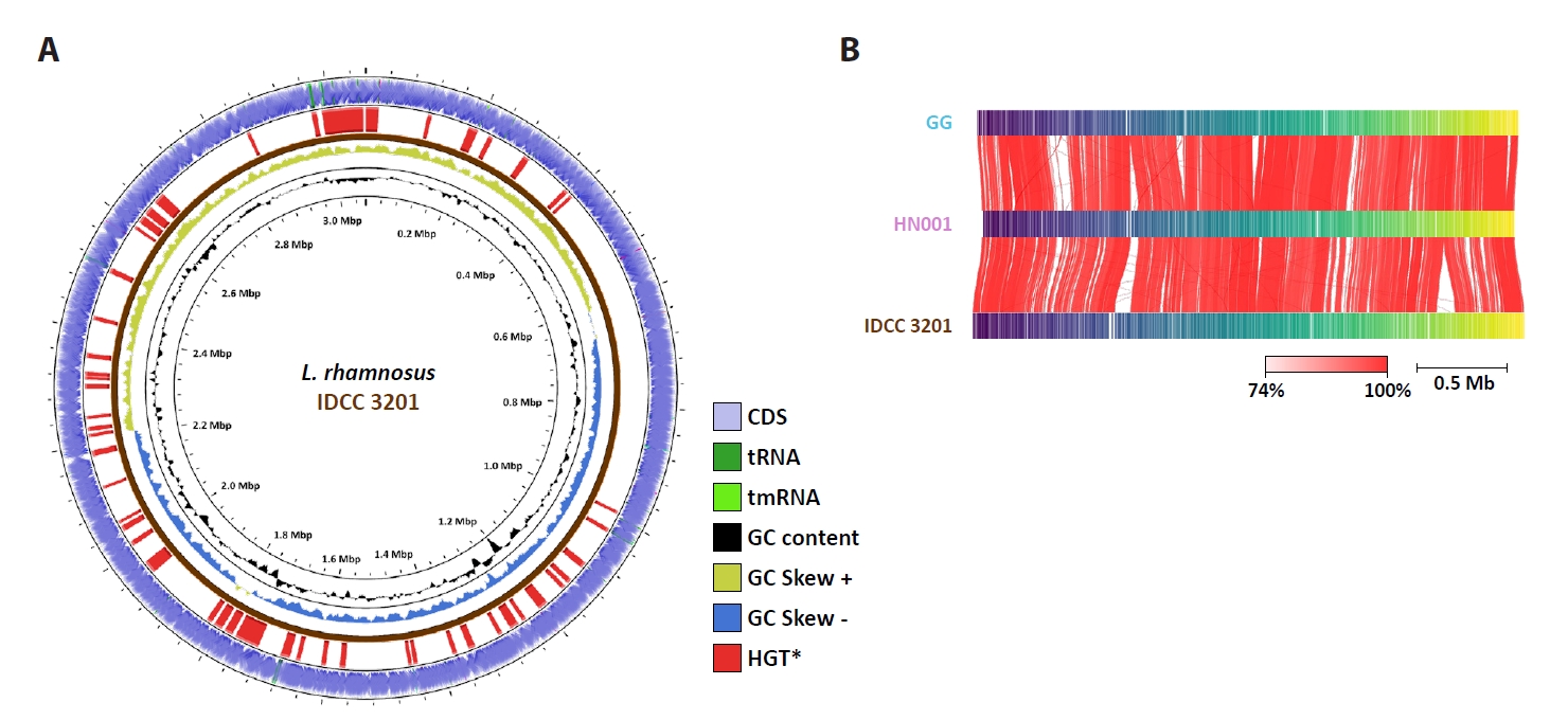


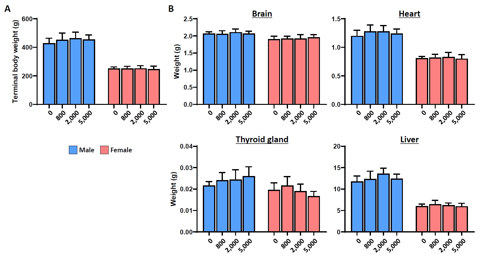
Fig. 1.
Fig. 2.
Fig. 3.
Fig. 4.
Fig. 5.
| Strains | IDCC 3201 | HN001 | GG |
|---|---|---|---|
| No. of contifgs | 1 | 2 | 1 |
| Plasmids | 0 | 1 | 0 |
| Genome size (bp |
3,064,263 | 2,992,974 | 3,010,111 |
| DNA G + C |
46.69 | 46.76 | 46.69 |
| No. of CDSs |
2,942 | 2,769 | 2,849 |
| No. of rRNA genes | 15 | 15 | 15 |
| No. of tRNA genes | 60 | 59 | 57 |
| Mean of CDS lengths (bp) | 836.9 | 903.1 | 851.2 |
| Median of CDS lengths (bp) | 717 | 786 | 738 |
| Mean of intergenic lengths (bp) | 137.2 | 154.9 | 131.4 |
| Median of intergenic lengths (bp) | 83 | 99 | 85 |
| Homology with IDCC 3201 by OrthoANI |
NA |
98.01% | 98.25% |
| Homology with IDCC 3201 by TNA |
NA | 99.85% | 99.82% |
| Strain | Em | Gm | Ap | Tc | Ca | Sm | Cm | Km | Vm |
|---|---|---|---|---|---|---|---|---|---|
| 1 | 16 | 4 | 8 | 4 | 32 | 1 | 64 | n.r.a | |
| 4 | 1 | 2b | 16 | 16 | 16 | - | 32 | 4b | |
| 1 | 128 | 4 | 4 | 16 | > 256 | 2 | > 256 | - | |
| 0.5 | 16 | 8 | 4 | - | 32 | 1 | - | - | |
| L. rhamnosus IDCC 3201 | 0.5 | 16 | 0.25 | 0.25 | 8 | 32 | 0.5 | 256 | > 256 |
| L. rhamnosus HN001 | 0.5 | 64 | 0.5 | 1 | 8 | 128 | 1 | 256 | > 256 |
| L. rhamnosus GGc | 0.25 | 0.25 | 1 | 2 | 8 | 64 | - | - | > 128 |
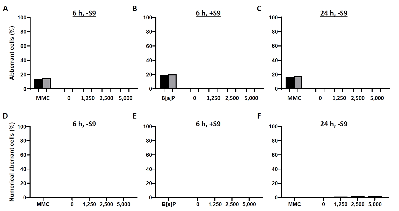
| Test item | Concentration (μg/ml) | RPD (%) | Percent of structural aberrant cells excluding gaps (%)a | Percent of structural aberrant cells including gaps (%)a | Percent of numerical aberrant cells (%)a |
|---|---|---|---|---|---|
| 6 h treatment without metabolic activation (6 h, -S9) | |||||
| RHT3201 | 0 | 100 | 0.667 | 1.000 | 0.000 |
| 1,250 | 97.1 | 0.333 | 0.333 | 0.333 | |
| 2,500 | 98.9 | 0.000 | 0.333 | 0.000 | |
| 5,000 | 74.2 | 0.333 | 0.667 | 0.667 | |
| MMC | 0.1 | 49.7 | 14.0* | 14.7 | 0.333 |
| 6 h treatment with metabolic activation (6 h, +S9) | |||||
| RHT3201 | 0 | 100 | 0.667 | 0.667 | 0.000 |
| 1,250 | 91.8 | 0.333 | 0.333 | 0.667 | |
| 2,500 | 92.4 | 0.000 | 0.000 | 0.333 | |
| 5,000 | 91.1 | 0.667 | 0.667 | 0.000 | |
| B[a]P | 20 | 48.8 | 19.0* | 20.0 | 0.667 |
| 24 h treatment without metabolic activation (24 h, -S9) | |||||
| RHT3201 | 0 | 100 | 0.333 | 1.000 | 0.667 |
| 1,250 | 94.7 | 0.000 | 0.000 | 1.000 | |
| 2,500 | 99.1 | 0.667 | 1.000 | 2.000 | |
| 5,000 | 90.6 | 0.000 | 0.000 | 2.000 | |
| MMC | 0.05 | 72.6 | 16.7* | 17.3 | 0.000 |
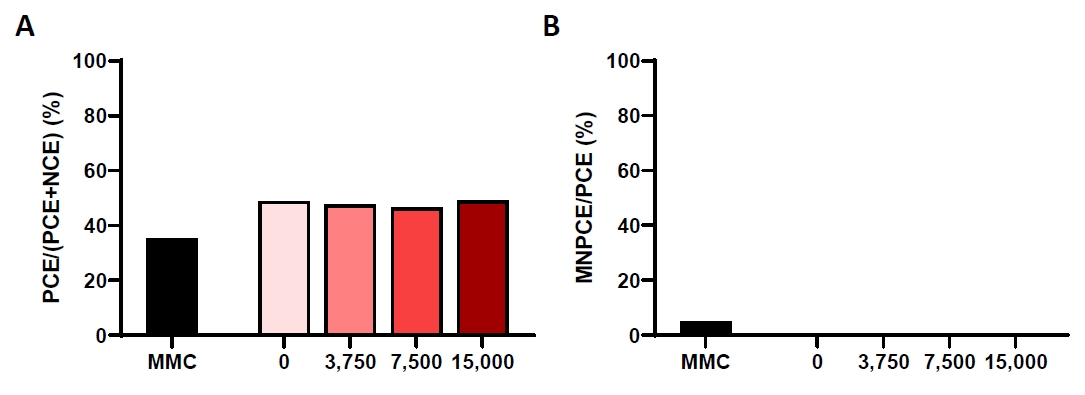
| Test item | Dose (mg/kg/day) | Percent of PCE/(PCE + NCE) (%) | Percent of MNPCE/PCE (%) |
|---|---|---|---|
| RHT3201 | 0 | 48.9 ± 0.91 | 0.075 ± 0.047 |
| 3,750 | 47.6 ± 3.69 | 0.070 ± 0.041 | |
| 7,500 | 46.5 ± 3.84 | 0.095 ± 0.057 | |
| 15,000 | 49.0 ± 3.77 | 0.055 ± 0.048 | |
| MMC | 2 | 35.2* ± 1.52 | 4.855* ± 0.359 |
| Dose (mg/kg/day) | Visual response | Touch response | Click response | Tail pinch response | Aerial righting reflex | Hindlimb landing foot splay (mm) | Forelimb grip strength (g) | Hindlimb grip strength (g) | ||||||
|---|---|---|---|---|---|---|---|---|---|---|---|---|---|---|
| Males | ||||||||||||||
| 0 | 3 ± 0 | 3 ± 0 | 3 ± 0 | 3 ± 0 | 0 ± 0 | 65.24 ± 18.33 | 1385 ± 112 | 800 ± 54 | ||||||
| 800 | 3 ± 0 | 3 ± 0 | 3 ± 0 | 3 ± 0 | 0 ± 0 | 68.98 ± 20.14 | 1414 ± 149 | 756 ± 113 | ||||||
| 2,000 | 3 ± 0 | 3 ± 0 | 3 ± 0 | 3 ± 0 | 0 ± 0 | 70.75 ± 18.17 | 1488 ± 174 | 762 ± 62 | ||||||
| 5,000 | 3 ± 0 | 3 ± 0 | 3 ± 0 | 3 ± 0 | 0 ± 0 | 66.74 ± 12.13 | 1459 ± 145 | 745 ± 86 | ||||||
| Dose (mg/kg/day) | Ambulatory counts (minutes interval) | |||||||||||||
| 0–10 | 10–20 | 20–30 | 30–40 | 40–50 | 50–60 | Total | ||||||||
| Males | ||||||||||||||
| 0 | 2020 ± 502 | 1156 ± 478 | 843 ± 669 | 526 ± 317 | 364 ± 316 | 437 ± 403 | 5346 ± 1770 | |||||||
| 800 | 1777* ± 376 | 1254 ± 436 | 897 ± 238 | 575 ± 328 | 424 ± 301 | 289 ± 312 | 5216 ± 1528 | |||||||
| 2,000 | 2399* ± 678 | 1422 ± 423 | 892 ± 417 | 758 ± 389 | 459 ± 564 | 327 ± 704 | 6256 ± 1936 | |||||||
| 5,000 | 2308 ± 572 | 1296 ± 477 | 673 ± 315 | 307 ± 335 | 157 ± 194 | 259 ± 286 | 5000 ± 1357 | |||||||
| Dose (mg/kg/day) | Vertical counts (minutes interval) | |||||||||||||
| 0–10 | 10–20 | 20–30 | 30–40 | 40–50 | 50–60 | Total | ||||||||
| Males | ||||||||||||||
| 0 | 115 ± 25 | 88 ± 30 | 67 ± 35 | 54 ± 32 | 49 ± 46 | 51 ± 42 | 424 ± 157 | |||||||
| 800 | 126 ± 36 | 98 ± 31 | 78 ± 31 | 56 ± 25 | 44 ± 23 | 31 ± 28 | 432 ± 124 | |||||||
| 2,000 | 108 ± 24 | 90 ± 22 | 59 ± 22 | 49 ± 23 | 34 ± 31 | 20 ± 31 | 359 ± 99 | |||||||
| 5,000 | 107 ± 43 | 72 ± 36 | 48 ± 26 | 33 ± 29 | 15 ± 19 | 16 ± 18 | 291 ± 128 | |||||||
| Dose (mg/kg/day) | Visual response | Touch response | Click response | Tail pinch response | Aerial righting reflex | Hindlimb landing foot splay (mm) | Forelimb grip strength (g) | Hindlimb grip strength (g) | ||||||
| Females | ||||||||||||||
| 0 | 3 ± 0 | 3 ± 0 | 3 ± 0 | 3 ± 0 | 0 ± 0 | 67.61 ± 12.21 | 1261 ± 140 | 607 ± 58 | ||||||
| 800 | 3 ± 0 | 3 ± 0 | 3 ± 0 | 3 ± 0 | 0 ± 0 | 61.66 ± 16.64 | 1277 ± 125 | 564 ± 89 | ||||||
| 2,000 | 3 ± 0 | 3 ± 0 | 3 ± 0 | 3 ± 0 | 0 ± 0 | 62.42 ± 16.30 | 1303 ± 116 | 627 ± 53 | ||||||
| 5,000 | 3 ± 0 | 3 ± 0 | 3 ± 0 | 3 ± 0 | 0 ± 0 | 62.14 ± 19.73 | 1218 ± 148 | 626 ± 67 | ||||||
| Dose (mg/kg/day) | Ambulatory counts (minutes interval) | |||||||||||||
| 0–10 | 10–20 | 20–30 | 30–40 | 40–50 | 50–60 | Total | ||||||||
| Females | ||||||||||||||
| 0 | 2559 ± 377 | 1496 ± 420 | 1221 ± 438 | 816 ± 432 | 542 ± 410 | 514 ± 304 | 7147 ± 1658 | |||||||
| 800 | 2370 ± 486 | 1498 ± 433 | 941 ± 239 | 568 ± 294 | 434 ± 358 | 530 ± 415 | 6342 ± 1174 | |||||||
| 2,000 | 2320 ± 478 | 1696 ± 448 | 1232 ± 405 | 937 ± 477 | 788 ± 567 | 478 ± 475 | 7451 ± 2223 | |||||||
| 5,000 | 2330 ± 623 | 1564 ± 440 | 950 ± 488 | 589 ± 422 | 641 ± 429 | 336 ± 306 | 6410 ± 1848 | |||||||
| Dose (mg/kg/day) | Vertical counts (minutes interval) | |||||||||||||
| 0–10 | 10–20 | 20–30 | 30–40 | 40–50 | 50–60 | Total | ||||||||
| Females | ||||||||||||||
| 0 | 110 ± 21 | 89 ± 28 | 64 ± 25 | 47 ± 31 | 35 ± 22 | 32 ± 22 | 378 ± 124 | |||||||
| 800 | 130 ± 19 | 93 ± 17 | 67 ± 22 | 51 ± 30 | 41 ± 28 | 32 ± 23 | 414 ± 106 | |||||||
| 2,000 | 121 ± 23 | 97 ± 22 | 73 ± 27 | 43 ± 22 | 55 ± 36 | 37 ± 30 | 426 ± 128 | |||||||
| 5,000 | 103 ± 41 | 70 ± 29 | 42 ± 25 | 28 ± 22 | 39 ± 36 | 23 ± 20 | 305 ± 150 | |||||||
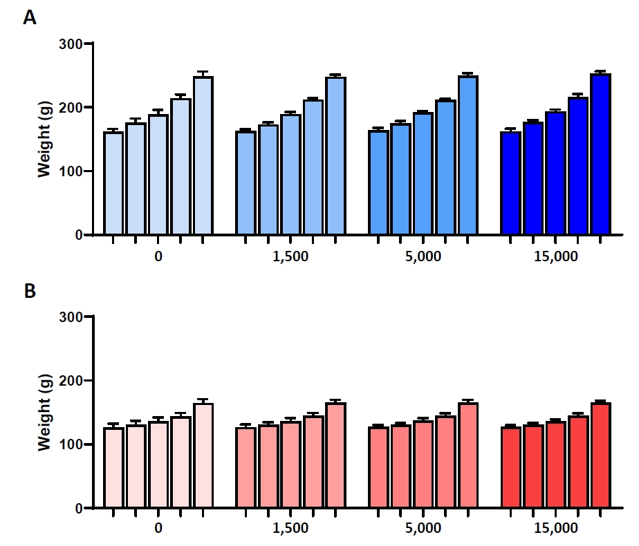
| Dose (mg/kg/day) | Terminal body weight (g) | Brain | Heart | Pituitary gland | ||||
|---|---|---|---|---|---|---|---|---|
| g | % | g | % | g | % | |||
| Males | ||||||||
| 0 | 429.3 ± 34.4 | 2.07 ± 0.05 | 0.49 ± 0.04 | 1.20 ± 0.10 | 0.28 ± 0.03 | 0.0127 ± 0.0013 | 0.0030 ± 0.0003 | |
| 800 | 452.3 ± 48.6 | 2.06 ± 0.09 | 0.46 ± 0.04 | 1.28 ± 0.11 | 0.29 ± 0.02 | 0.0136 ± 0.0007 | 0.0031 ± 0.0003 | |
| 2,000 | 463.0 ± 44.0 | 2.11 ± 0.09 | 0.46 ± 0.03 | 1.28 ± 0.10 | 0.28 ± 0.02 | 0.0131 ± 0.0016 | 0.0028 ± 0.0002 | |
| 5,000 | 455.3 ± 31.8 | 2.07 ± 0.07 | 0.45 ± 0.04 | 1.24 ± 0.08 | 0.27 ± 0.02 | 0.0132 ± 0.0013 | 0.0029 ± 0.0003 | |
| Dose (mg/kg/day) | Liver | Spleen | Lung | Thyroid gland |
||||
| g | % | g | % | g | % | g | % | |
| Males | ||||||||
| 0 | 11.79 ± 1.27 | 2.75 ± 0.21 | 0.76 ± 0.12 | 0.18 ± 0.05 | 1.59 ± 0.12 | 0.37 ± 0.03 | 0.0216 ± 0.0019 | 0.0051 ± 0.0005 |
| 800 | 12.34 ± 1.85 | 2.73 ± 0.27 | 0.75 ± 0.12 | 0.17 ± 0.02 | 1.65 ± 0.14 | 0.36 ± 0.02 | 0.0241 ± 0.0036 | 0.0054 ± 0.0010 |
| 2,000 | 13.60 |
2.95 ± 0.22 | 0.82 ± 0.15 | 0.18 ± 0.02 | 1.67 ± 0.18 | 0.36 ± 0.02 | 0.0244 ± 0.0046 | 0.0053 ± 0.0009 |
| 5,000 | 12.46 ± 1.07 | 2.74 ± 0.20 | 0.75 ± 0.08 | 0.16 ± 0.01 | 1.65 ± 0.09 | 0.36 ± 0.02 | 0.0260 ± 0.0044 | 0.0057 ± 0.0007 |
| Dose (mg/kg/day) | Kidney | Thymus | Testis | Adrenal gland | ||||
| g | % | g | % | g | % | g | % | |
| Males | ||||||||
| 0 | 2.69 ± 0.25 | 0.63 ± 0.05 | 0.30 ± 0.06 | 0.07 ± 0.01 | 3.79 ± 0.26 | 0.88 ± 0.06 | 0.0462 ± 0.0033 | 0.0108 ± 0.0011 |
| 800 | 2.74 ± 0.24 | 0.61 ± 0.05 | 0.30 ± 0.06 | 0.07 ± 0.01 | 4.00 ± 0.15 | 0.89 ± 0.09 | 0.0533 |
0.0118 ± 0.0017 |
| 2,000 | 2.87 ± 0.22 | 0.62 ± 0.05 | 0.31 ± 0.09 | 0.07 ± 0.02 | 3.90 ± 0.37 | 0.85 ± 0.08 | 0.0494 ± 0.0070 | 0.0107 ± 0.0012 |
| 5,000 | 2.80 ± 0.24 | 0.62 ± 0.05 | 0.32 ± 0.09 | 0.07 ± 0.02 | 3.99 ± 0.24 | 0.88 ± 0.05 | 0.0510 ± 0.0134 | 0.0111 ± 0.0025 |
| Dose (mg/kg/day) | Epididymis | Prostate |
||||||
| g | % | g | % | |||||
| Males | ||||||||
| 0 | 1.37 ± 0.16 | 0.32 ± 0.04 | 3.18 ± 0.38 | 0.74 ± 0.09 | ||||
| 800 | 1.46 ± 0.08 | 0.32 ± 0.03 | 3.27 ± 0.50 | 0.72 ± 0.09 | ||||
| 2,000 | 1.44 ± 0.17 | 0.31 ± 0.03 | 3.23 ± 0.29 | 0.70 ± 0.07 | ||||
| 5,000 | 1.43 ± 0.13 | 0.31 ± 0.01 | 3.18 ± 0.53 | 0.70 ± 0.10 | ||||
| Dose (mg/kg/day) | Terminal body weight (g) | Brain | Heart | Pituitary gland | ||||
| g | % | g | % | g | % | |||
| Females | ||||||||
| 0 | 251.0 ± 11.7 | 1.90 ± 0.09 | 0.76 ± 0.06 | 0.81 ± 0.03 | 0.32 ± 0.02 | 0.0158 ± 0.0021 | 0.0063 ± 0.0010 | |
| 800 | 250.6 ± 16.7 | 1.92 ± 0.07 | 0.77 ± 0.05 | 0.82 ± 0.06 | 0.33 ± 0.02 | 0.0166 ± 0.0029 | 0.0067 ± 0.0013 | |
| 2,000 | 252.2 ± 19.0 | 1.92 ± 0.12 | 0.76 ± 0.05 | 0.83 ± 0.08 | 0.33 ± 0.02 | 0.0169 ± 0.0029 | 0.0067 ± 0.0010 | |
| 5,000 | 246.2 ± 22.4 | 1.96 ± 0.08 | 0.80 ± 0.06 | 0.80 ± 0.07 | 0.33 ± 0.01 | 0.0155 ± 0.0019 | 0.0063 ± 0.0007 | |
| Dose (mg/kg/day) | Liver | Spleen | Lung | Thyroid gland |
||||
| g | % | g | % | g | % | g | % | |
| Females | ||||||||
| 0 | 6.02 ± 0.48 | 2.40 ± 0.15 | 0.55 ± 0.08 | 0.22 ± 0.03 | 1.19 ± 0.06 | 0.47 ± 0.03 | 0.0196 ± 0.0033 | 0.0078 ± 0.0015 |
| 800 | 6.48 ± 0.88 | 2.58 ± 0.28 | 0.56 ± 0.08 | 0.22 ± 0.02 | 1.21 ± 0.09 | 0.48 ± 0.02 | 0.0216 ± 0.0042 | 0.0086 ± 0.0014 |
| 2,000 | 6.24 ± 0.52 | 2.48 ± 0.18 | 0.60 ± 0.11 | 0.24 ± 0.04 | 1.24 ± 0.13 | 0.49 ± 0.03 | 0.0190 ± 0.0033 | 0.0076 ± 0.0014 |
| 5,000 | 5.98 ± 0.70 | 2.42 ± 0.15 | 0.54 ± 0.04 | 0.22 ± 0.02 | 1.20 ± 0.09 | 0.49 ± 0.02 | 0.0167 ± 0.0022 | 0.0068 ± 0.0008 |
| Dose (mg/kg/day) | Kidney | Thymus | Uterus | Adrenal gland | ||||
| g | % | g | % | g | % | g | % | |
| Females | ||||||||
| 0 | 1.61 ± 0.11 | 0.64 ± 0.03 | 0.23 ± 0.05 | 0.09 ± 0.02 | 0.62 ± 0.26 | 0.24 ± 0.11 | 0.0624 ± 0.0107 | 0.0249 ± 0.0044 |
| 800 | 1.65 ± 0.11 | 0.66 ± 0.05 | 0.22 ± 0.04 | 0.09 ± 0.01 | 0.65 ± 0.17 | 0.26 ± 0.07 | 0.0663 ± 0.0082 | 0.0266 ± 0.0039 |
| 2,000 | 1.68 ± 0.16 | 0.67 ± 0.06 | 0.23 ± 0.08 | 0.09 ± 0.02 | 0.67 ± 0.23 | 0.26 ± 0.08 | 0.0699 ± 0.0071 | 0.0279 ± 0.0036 |
| 5,000 | 1.65 ± 0.20 | 0.67 ± 0.06 | 0.23 ± 0.03 | 0.09 ± 0.01 | 0.73 ± 0.24 | 0.30 ± 0.10 | 0.0636 ± 0.0087 | 0.0258 ± 0.0020 |
| Dose (mg/kg/day) | Ovary | |||||||
| g | % | |||||||
| Females | ||||||||
| 0 | 0.0818 ± 0.0105 | 0.0326 ± 0.0040 | ||||||
| 800 | 0.0828 ± 0.0115 | 0.0330 ± 0.0039 | ||||||
| 2,000 | 0.0915 ± 0.0188 | 0.0362 ± 0.0064 | ||||||
| 5,000 | 0.0869 ± 0.0181 | 0.0351 ± 0.0057 | ||||||
abp = base pair; bG + C = guanine + cytosine; cCDSs = coding sequences; dANI = average nucleotide identity; eTNA = tetra-nucleotide analysis; fNA = not applicable.
Values are Mean. Em, erythromycin; Gm, gentamicin; Ap, ampicillin; Tc, tetracycline; Ca, chloramphenicol; Sm, streptomycin; Cm, clindamycin; Km, kanamycin; Vm, vancomycin. an.r., not required; bNote by SCAN - Certain species are inherently resistant; c
Values are Mean. MMC, mitomycin C; B[a]P, benzo[a]pyrene; RPD, relative population doubling. aMean percentage of duplicate culture; total 300 metaphase cells were examined (150 cells/culture). *Significantly different from the vehicle control at
Values are Mean ± SD. MMC, mitomycin C; PCE, polychromatic erythrocyte; NCE, normochromatic erythrocyte; MNPCE, micronucleated polychromatic erythrocyte. *Significantly different from the vehicle control at
Values are Mean ± SD. Visual response 3: the animal approaches slowly and smells a stimulating bar; Touch response 3: the animal turns around slowly; Click response 3: twitching of body; Tail pinch response 3: squeaking, turning back; Aerial righting reflex 0: normal (landing on four limbs). *Significantly different from the vehicle control at
Values are Mean ± SD. Organ weight for thyroid gland with parathyroid gland as a whole. Organ weight for prostate with seminal vesicle and coagulation gland as a whole. Significantly different from the vehicle control at
Table 1.
Table 2.
Table 3.
Table 4.
Table 5.
Table 6.
TOP

