Skip Navigation
Skip to contents

Journal of Microbiology : Journal of Microbiology

OPEN ACCESS
SEARCH
Search

Articles

Page Path
HOME > J. Microbiol > Ahead of print > Article
Review
Influenza A viruses in bats: Viral characteristics and zoonotic risk at the wildlife-human interface
Kwang-Min Yu1, Su-Jin Park1,2,*orcid

DOI: https://doi.org/10.71150/jm.2608013
Published online: November 10, 2026

1Research Institute of Molecular Alchemy (RIMA), Gyeongsang National University, Jinju 52828, Republic of Korea

2Division of Life Science, College of Natural Sciences, Gyeongsang National University, Jinju 52828, Republic of Korea

*Correspondence Su-Jin Park parksujin@gnu.ac.kr
This is an Open Access article distributed under the terms of the Creative Commons Attribution 4.0 International License (CC BY 4.0) (https://creativecommons.org/licenses/by/4.0/) which permits unrestricted use, distribution, and reproduction in any medium, provided the original work is properly cited.
• Received: August 14, 2026   • Revised: September 22, 2026   • Accepted: September 28, 2026

© 2026 The Authors. Published under licence by The Microbiological Society of Korea.

This is an Open Access article distributed under the terms of the Creative Commons Attribution 4.0 International License (CC BY 4.0) (https://creativecommons.org/licenses/by/4.0/) which permits unrestricted use, distribution, and reproduction in any medium, provided the original work is properly cited.

  • 35 Views
  • 1 Download
  • The discovery of influenza A viruses in bats has changed our understanding of viral circulation in nature. For years, wild aquatic birds were considered the primary natural reservoir for influenza A viruses. However, surveillance efforts in Neotropical bats have revealed highly divergent bat-derived lineages, including H17N10, H18N11, and H18N12. Researchers also isolated an avian-like H9N2 virus from Egyptian fruit bats. These findings show that bats do not carry a single influenza virus type; instead, they are associated with multiple lineages with distinct evolutionary backgrounds. The H17 and H18 viruses appear to be adapted to bat physiology. They infect cells via major histocompatibility complex class II (MHC-II) molecules rather than the usual sialic acid receptors; their neuraminidase-like proteins lack conventional sialidase activity; and they replicate poorly in non-bat mammalian models. In contrast, the bat-associated, avian-like H9N2 virus behaves differently. Although its surface proteins retain avian-like characteristics, it replicates efficiently and transmits readily in mammalian systems, together with reduced sensitivity to human MxA restriction. These observations raise the possibility that bats could act as ecological bridging hosts for avian influenza viruses. However, there is currently no direct field evidence that avian influenza viruses acquire mammalian-compatible traits in bats or are subsequently transmitted from bats to other host species. This review provides an overview of the current understanding of bat-associated influenza viruses and their potential implications for global health.
With over 1,400 recognized species, bats represent approximately 20% of all mammalian species and are distributed worldwide, making them remarkable natural reservoirs for diverse zoonotic pathogens (Ingala et al., 2018; Olival et al., 2017). Beyond their established role in harboring coronaviruses, filoviruses, lyssaviruses, and paramyxoviruses, bats have emerged as unexpected reservoirs for phylogenetically distinct influenza A viruses (IAVs), fundamentally expanding understanding of viral ecology and the host range of influenza (Banerjee et al., 2019; Banyard et al., 2011; CDC, 2024; Drexler et al., 2012; Leroy et al., 2005; Li et al., 2005; Osman et al., 2025; Tong et al., 2012, 2013). The discovery of highly divergent influenza A-like viruses in bat populations in 2012 revealed that bats harbor complex viral populations with evolutionary trajectories distinct from those of classical avian and mammalian IAV lineages (Tong et al., 2012).
IAVs possess an eight-segmented negative-sense RNA genome (McCauley et al., 2009). The viruses are classified according to the antigenic properties of their two surface glycoproteins: hemagglutinin (HA) and neuraminidase (NA). Wild aquatic birds are considered the primary natural reservoirs, harboring most known influenza A virus subtypes, including H1–H16 and H19 HA, as well as N1–N9 NA (Fereidouni et al., 2023; Horimoto and Kawaoka, 2005; Karakus et al., 2024). These birds facilitate transmission of IAVs to a wide range of avian and mammalian species. Host susceptibility is largely determined by the preference of the HA protein for either α2,3-linked (avian type) or α2,6-linked (mammalian type) sialic acid receptors, while genetic reassortment between co-infecting viruses often drives the emergence of novel strains.
The first evidence of bat-derived influenza lineages came with the identification of H17N10 in northern yellow-shouldered bats (Sturnira parvidens) and H18N11 in flat-faced fruit-eating bats (Artibeus planirostris) from Central and South America sampled in 2009–2010 (Tong et al., 2012, 2013). H18N12, tentatively proposed as a new subtype, was later reported in a pool of lesser bulldog bats (Noctilio albiventris) (Echeverri-De la Hoz et al., 2025). These lineages are genetically distinct from conventional avian and mammalian IAVs and are referred to as bat-derived because they have been detected exclusively in bats to date. Molecular and serological evidence, particularly for H17N10 and H18N11, indicates exposure and possible circulation in bat populations, although sustained natural maintenance has not been established through longitudinal studies.
A distinct bat-associated, avian-like H9N2 virus was isolated from Egyptian fruit bats (Rousettus aegyptiacus), and a closely related virus was subsequently detected in South Africa (Kandeil et al., 2019; Rademan et al., 2023). Unlike the deeply divergent H17/H18 lineages, these bat-associated H9N2 viruses are genetically similar to classical avian IAVs, and the Egyptian isolate can be propagated in embryonated chicken eggs. Under experimental conditions, R. aegyptiacus is susceptible to the bat-associated isolate but not to the conventional avian H9N2 strains tested (Halwe et al., 2021). The detections at two geographically separated locations indicate that the lineage is not confined to a single site, but sustained natural transmission within bat populations has not yet been demonstrated.
To our knowledge, previous reviews have focused primarily on the divergent H17 and H18 lineages, whereas reviews covering both bat-derived H17/H18 and bat-associated H9N2 have generally discussed them separately by subtype rather than comparing them within a common framework (Ciminski and Schwemmle, 2021; Ciminski et al., 2017; Ma et al., 2015; Yang et al., 2021). Recent studies have provided comparative data on receptor usage, replication in experimental models, host range, and transmission, but these findings have not been integrated systematically across the two groups (Griffiths et al., 2025; Halwe et al., 2024; Wang et al., 2024).
For this narrative review, we searched PubMed for articles published between 2012 and September 2026 using combinations of the search terms 'bat', 'influenza A virus', 'H17N10', 'H18N11', 'H18N12', 'H9N2', and 'spillover'. We included peer-reviewed original studies, surveillance reports, and reviews addressing the genomic, structural, ecological, or biological characteristics of bat-associated influenza viruses. Studies of non-bat IAVs were included only when they provided a direct comparison, and conference abstracts without supporting data were excluded.
Here, we compare the bat-derived H17/H18 lineages with the bat-associated H9N2 lineage in receptor usage and entry, polymerase host-range determinants, innate immune restriction, genome-packaging compatibility, and surveillance data (Tables 1 and S1). We also reassess their potential zoonotic significance by distinguishing phenotypes observed in experimental systems from field evidence of natural maintenance in bat populations.
Basic biological characteristics of bats contribute to their ability to maintain and spread viruses (Gonzalez and Banerjee, 2022; Gorbunova et al., 2020). These include living in large, densely populated colonies, long lifespans relative to their body size, and powered flight over long distances (Calisher et al., 2006; Luis et al., 2013). These traits increase the risk of viral exposure within roosts, facilitate the long-term persistence of pathogens, and enable the spread of viral populations across large geographic areas (Luis et al., 2013). Host–virus coevolution may have influenced the diversification of bat-associated viruses, as bat immune genes show more widespread signatures of positive selection than those of other mammalian orders (Morales et al., 2025). Some bat species also exhibit distinctive antiviral and inflammatory responses that may promote tolerance to certain viral infections and limit clinical disease (Irving et al., 2021).
Although bat immunology remains limited, some bat species exhibit an attenuated innate inflammatory response despite sharing immune cell functions with humans and other mammals (Irving et al., 2021). This attenuated response in some bat species may have led to the selection of viruses that are highly adapted to bat physiology and, hence, fundamentally different from those circulating in birds and other mammals. Therefore, the study of bat-associated lineages will further deepen understanding of the limits of viral evolutionary plasticity and the constraints that influence viral host adaptation.
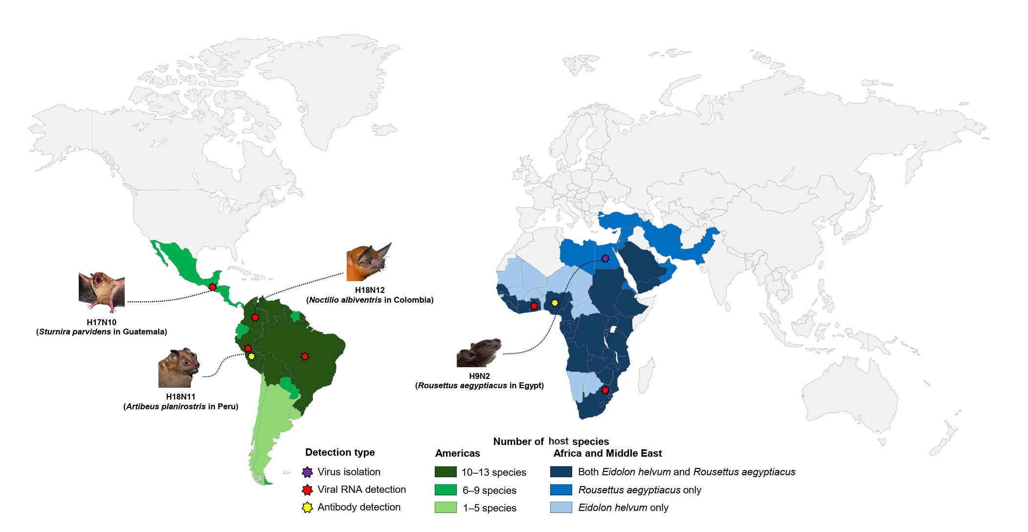
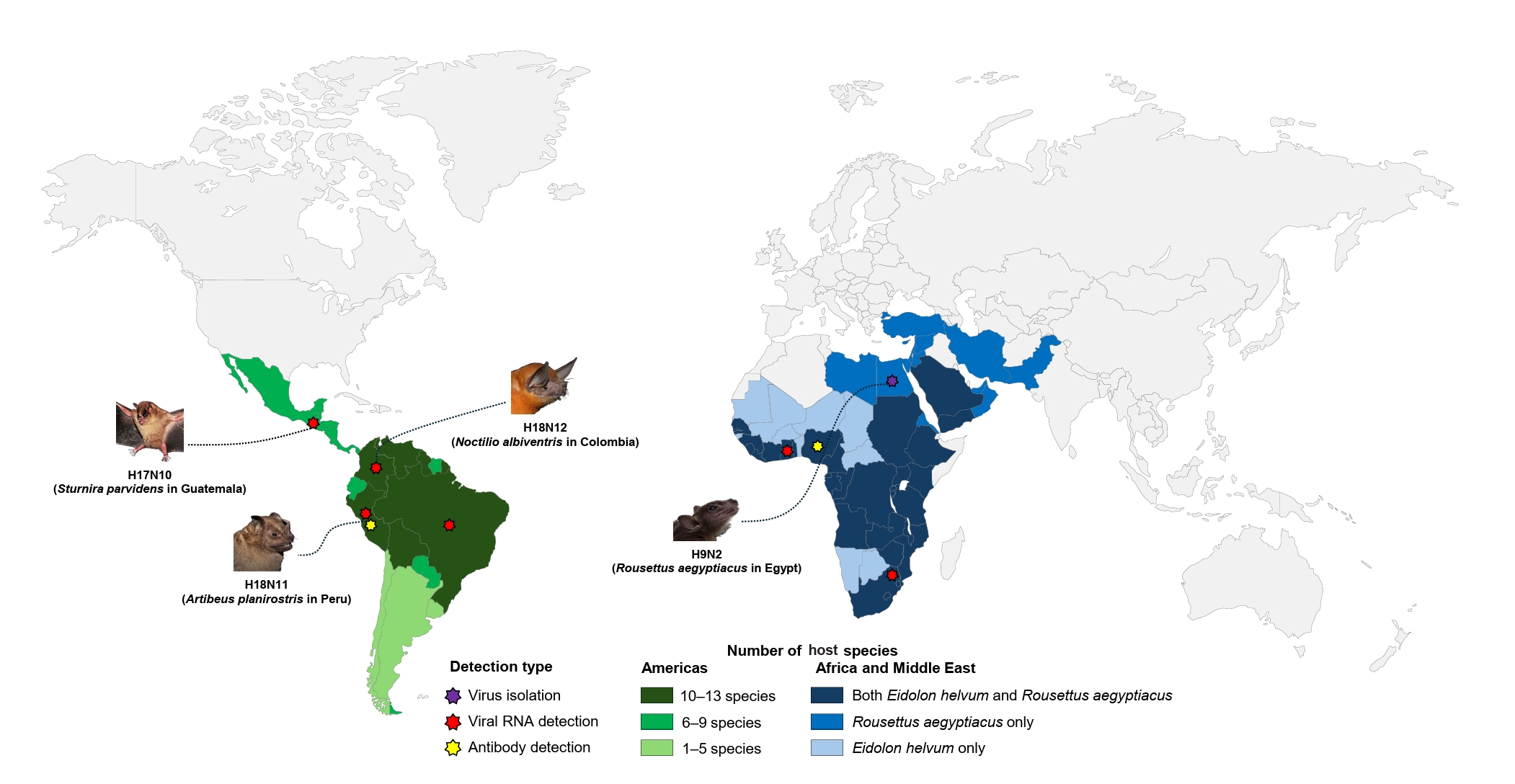
Bat-associated influenza viruses have been detected in species occupying diverse ecological niches across multiple continents (Campos et al., 2019; Echeverri-De la Hoz et al., 2025; Kandeil et al., 2019; Rademan et al., 2023; Tong et al., 2012, 2013). Figure 1 shows the country-level distributions of the bat species from which these viruses have been reported, together with the locations of reported field findings; the underlying evidence for each finding is listed in Table S1.
In the Neotropics, hosts of H17N10 and H18N11/H18N12 are widely distributed across Central and South America, including landscapes influenced by human agricultural activities (Campos et al., 2019; Echeverri-De la Hoz et al., 2025; Tong et al., 2012, 2013). As shown in Fig. 1, countries such as Peru and Brazil contain multiple reported host species, including the great fruit-eating bat (A. lituratus) and Seba's short-tailed bat (Carollia perspicillata), as well as the common vampire bat (Desmodus rotundus), a blood-feeding species. Their differing feeding and roosting habits may influence opportunities for contact and exposure within agricultural landscapes. In Africa, R. aegyptiacus frequently roosts in caves and human-made structures, which may create opportunities for contact with domestic animals and humans (Geldenhuys et al., 2023). The country-level distributions of R. aegyptiacus and Eidolon helvum overlap, although it remains unclear whether these species share local habitats or encounter common sources of exposure. Such zones of co-occurrence therefore represent priorities for targeted surveillance, although co-occurrence at the country level does not by itself imply shared roosting sites, direct interspecies contact, or cross-species transmission.
Field studies have detected H18N11 RNA in rectal swabs and intestinal tissue, but RNA detection alone does not establish active replication or tissue tropism (Campos et al., 2019; Tong et al., 2013). Intestinal replication and fecal shedding were subsequently observed in experimentally infected Jamaican fruit bats (A. jamaicensis) (Ciminski et al., 2019b; Kessler et al., 2024). These experimental findings should be distinguished from field observations and do not, by themselves, establish environmental transmission through contaminated soil or water.
In Africa, bat-associated H9N2 has been isolated from R. aegyptiacus in Egypt, and viral RNA has subsequently been detected in the same species in South Africa (Kandeil et al., 2019; Rademan et al., 2023). In contrast, evidence from the straw-coloured fruit bat (E. helvum) in Ghana is limited to serological reactivity against influenza antigens, including H9 haemagglutinin, and does not establish infection with the bat-associated H9N2 lineage (Freidl et al., 2015).
In Peru, antibodies to recombinant H18 HA or N11 were detected in 55 of 110 bats, whereas viral RNA was detected in only one animal (Tong et al., 2013). This discrepancy between low molecular detection rates and high seroprevalence poses challenges for interpreting surveillance data (Table S1). Seropositivity indicates previous exposure or antigenic reactivity but does not indicate active infection, and cross-reactivity with related antigens cannot always be excluded (Freidl et al., 2015). This pattern underscores the need for integrated surveillance combining molecular and serological evidence to distinguish current infection from historical exposure.
A long-term study of D. rotundus documented sustained H18 seropositivity, consistent with endemic circulation, while mechanistic modeling supported a possible maintenance role for vampire bats rather than fruit bats (Griffiths et al., 2025). These findings do not provide direct virological confirmation of sustained circulation. Considered alongside the experimental evidence of intestinal replication and fecal shedding, the serological and modeling findings support surveillance that accounts for non-respiratory infection and examines D. rotundus as a host of interest.
Bat-associated influenza viruses fall into two broad evolutionary categories: the highly divergent bat-derived lineages (H17N10, H18N11, and H18N12) and the bat-associated H9N2 lineage (Campos et al., 2019; Kandeil et al., 2019; Rademan et al., 2023; Tong et al., 2012, 2013). Phylogenetic analyses show that the H17 and H18 HAs belong to Group 1 HAs but form a deeply divergent monophyletic clade. H17 shares approximately 45% amino acid identity with conventional IAV HAs, whereas H18 has a mean pairwise identity of 49.1% with Group 1 HAs (Tong et al., 2012, 2013; Zhu et al., 2013). Their NA-like proteins (N10–N12) are even more divergent, with N10 and N11 sharing 20–27% and approximately 29.6% amino acid identity, respectively (Li et al., 2012; Tong et al., 2013; Zhu et al., 2012).
A SuperNetwork constructed from the six internal gene segments (PB2, PB1, PA, NP, M, and NS) of conventional and bat-derived IAVs placed the H17 and H18 segments as basal outgroups to conventional IAVs (Ciminski et al., 2020). Nucleotide divergence between H17/H18 and H1–H16 viruses exceeds 25% across most gene segments and approaches 40% for PB2 and PA (Volkhin et al., 2026). In a Bayesian time-calibrated phylogeny of PB1 inferred under a relaxed molecular clock, the H17 and H18 lineage was estimated to have diverged more than 650 years ago (Ciminski et al., 2020). In addition, the NA-like segment of the proposed N12 subtype was found to occupy a basal position relative to N11 and was associated with a node dated to the mid-20th century, suggesting that it did not descend directly from N11 (Echeverri-De la Hoz et al., 2025). These dates are approximate because they depend on the evolutionary models applied to sequence data collected from limited geographic areas, host species, and sampling periods. Rates estimated from a narrow range of sampling dates may also be unreliable when extrapolated to nodes several centuries old. Despite this uncertainty, tree topology and genome divergence support the long-term independent evolution of the H17N10, H18N11, and H18N12 lineages in bats.
In contrast, bat-associated H9N2 has a closer and apparently more recent evolutionary relationship with conventional IAVs. In the same time-calibrated PB1 phylogeny, its PB1 lineage shared a most recent common ancestor with conventional IAVs at approximately 1,700 CE, whereas bat-associated H9 HA is closely related to more recent avian H9 sequences. The distinct evolutionary histories of PB1 and HA may reflect reassortment or the absence of ancestral intermediates, although the underlying evolutionary events remain unresolved (Ciminski et al., 2020; Halwe et al., 2024). Consistent with this closer relationship, the gene segments of bat-associated H9N2 viruses share approximately 72–87% nucleotide identity with those of conventional IAVs (Kandeil et al., 2019).
Despite these differences in evolutionary depth, the direction of host transition remains uncertain. Ciminski et al. (2020) considered both an ancestral bat origin of present-day IAVs and the introduction of an older avian precursor into mammals to be possible. Karamendin et al. (2024) dated the divergence between the bat and avian H9 lineages to the end of the 18th century and interpreted the phylogenetic evidence as supporting wild birds as the likely source of introduction into bats. These molecular-clock estimates and phylogenetic interpretations are model-dependent and constrained by sparse geographic and host sampling. The timing of the host transition, therefore, remains uncertain. The available phylogenetic evidence favors bird-to-bat transmission, but this interpretation should not be treated as a definitive conclusion. Compared with the H17/H18 lineages, the bat-associated H9N2 lineage is more closely related to conventional IAVs and appears to have diverged more recently. Available sequences support divergence between the bat-associated and avian H9N2 lineages, but whether this divergence represents functional adaptation to bats requires experimental investigation.
Surface glycoproteins evolution and function
The evolutionary divergence between the surface glycoproteins of bat-derived H17/18 and bat-associated H9 influenza viruses demonstrates substantial adaptation. Although H17 and H18 HAs have the characteristic trimeric structure of influenza HAs, their receptor-binding sites have undergone substantial remodeling through multiple amino acid substitutions and structural rearrangements (Sun et al., 2013; Tong et al., 2013). In classical HAs, the receptor-binding site forms a shallow pocket that coordinates sialic acid through conserved residues (Eisen et al., 1997; Martín et al., 1998; Skehel and Wiley, 2000; Weis et al., 1988; Wiley and Skehel, 1987; Zhu et al., 2013).
In the H17 and H18 variants, the canonical receptor-binding pocket is structurally remodeled and does not recognize conventional α2,3- or α2,6-linked sialic acids (Sun et al., 2013; Zhu et al., 2013). Early recombinant-protein studies did not detect the classical low-pH-induced conformational change in H17 HA, although subsequent pseudotype experiments showed that H17/H18-mediated entry depends on endosomal acidification (Hoffmann et al., 2016; Zhu et al., 2013). Moreover, the acid-stable H18 HA does not require M2-mediated preservation of its metastable conformation (Thompson et al., 2023). H17 and H18 instead use major histocompatibility complex class II (MHC-II) molecules for entry, with cell susceptibility depending partly on MHC-II expression and additional cellular determinants (Giotis et al., 2019; Karakus et al., 2019; Osman et al., 2025). Proteolytic activation by transmembrane serine proteases such as TMPRSS2 enables H17/H18 HA-mediated entry. However, in the cell systems tested, host-range restriction occurred primarily at the level of receptor engagement rather than proteolytic activation or low-pH triggering (Hoffmann et al., 2016).
The NA-like proteins (N10 and N11) form tetrameric structures similar to classical NAs but crucially lack key residues required for sialidase activity; the proposed N12 has been characterized only by homology modeling (Echeverri-De la Hoz et al., 2025; Li et al., 2012; Tong et al., 2013; Zhu et al., 2012). In this regard, it has been proposed that N11 may downregulate MHC-II expression to facilitate virion release, and it would represent an alternative function for N11 compared with classical NAs (Ciminski et al., 2019b). Mutations arising during serial passage in MDCK II cells enhanced N11-mediated viral replication, suggesting broader roles in the viral life cycle that remain incompletely understood (Zhong et al., 2020). In contrast, the bat-associated H9N2 retains typical avian-like glycoprotein functions, with conserved receptor-binding sites consistent with a preference for α2,3-linked sialic acids and catalytic NA activity, suggesting conventional sialic acid-dependent entry and release mechanisms (Halwe et al., 2024; Kandeil et al., 2019).
Molecular determinants of host range in the polymerase complex
The viral polymerase complex has been described as a major host-range determinant (Long et al., 2016; Subbarao et al., 1993). A single substitution at residue 282 in the PB2 subunit distinguishes bat H17 and H18 viruses from conventional IAV strains. The H17 and H18 viruses typically carry serine (S) at position 282, whereas avian and human IAVs carry glutamic acid (E) (Banerjee et al., 2022). Introducing the PB2-E282S substitution into a conventional IAV polymerase reduced polymerase activity in mammalian cells, whereas the reciprocal PB2-S282E substitution increased the activity of the bat virus polymerase in human cells. In the bat polymerase, the mammalian-adaptive PB2-S627K substitution also increased activity, but combining it with S282E gave only a minor increase, indicating that the two residues do not act cumulatively (Banerjee et al., 2022).
ANP32A does not appear to be the limiting factor for this restriction, since overexpression of chicken or human ANP32A rescued the PB2-627 phenotype but not the 282 phenotype (Banerjee et al., 2022). ANP32 proteins are nevertheless the best-characterized host cofactors of the influenza polymerase, and their usage differs both between species and among family members (Long et al., 2016, 2019; Park et al., 2020; Peacock et al., 2020, 2023). Bat ANP32A and ANP32B, like other mammalian orthologs, lack the avian-specific 33-amino-acid insertion and poorly support non-adapted avian polymerases (Long et al., 2016; Peacock et al., 2020). Notably, these H17/H18 polymerases instead rely most strongly on mammalian ANP32B, which supports them better than either bat ANP32 protein (Peacock et al., 2020), indicating that ANP32 usage in bat-derived viruses is not predictable from host species alone. The ANP32 cofactor usage of the bat-associated H9N2 lineage has not been directly characterized, representing an important gap in our understanding of its host adaptation. Importantly, its PB2 retains the avian-type glutamic acid at position 627 while carrying asparagine at position 701 (Kandeil et al., 2019). Although avian-type PB2 627E polymerases are generally restricted by mammalian ANP32 proteins, D701N independently adapts the polymerase to use them and, unlike E627K, is not strongly biased toward ANP32B, consistent with its emergence in hosts where ANP32A is the dominant pro-viral paralog (Peacock et al., 2023). The presence of D701N in this lineage therefore raises the possibility that it confers at least partial compatibility with mammalian-type ANP32 proteins, although this has not been tested for the bat virus polymerase. Minigenome assays in ANP32A/B-deficient cells complemented with chicken, bat, or human ANP32A and ANP32B, combined with reciprocal 701N/701D substitutions in a common backbone, would help determine whether this lineage has acquired this ability and whether its paralog preference differs among mammalian hosts.
Host-range restriction also occurs at the level of innate immunity. Human-adapted IAVs overcome restriction by human MxA through adaptive substitutions in the nucleoprotein, which are largely absent from avian strains (Mänz et al., 2013). Bat Mx1, the ortholog of human MxA, restricted H17N10, with Mx1 of C. perspicillata inhibiting virus-like particle formation as effectively as human MxA (Fuchs et al., 2017). H18N11 NP similarly lacks these escape substitutions, and although it confers partial resistance to both proteins, a chimeric PR8-H18N11 virus remained strongly restricted in MxA-expressing cells and in hMxA-transgenic mice (Ciminski et al., 2019a). The bat-associated H9N2, in contrast, carries one such substitution, methionine at position 316 in NP, yet still shows reduced replication in the presence of human MxA, suggesting that full resistance requires additional changes (Halwe et al., 2024).
Other internal genes (M and NS)
While HA and NA proteins often determine receptor specificity and initial host entry, the internal gene segments of bat-associated influenza viruses pose an additional critical barrier to reassortment and cross-species transmission. These incompatibilities limit the formation of reassortant viruses capable of efficient replication in new hosts.
The matrix (M) and non-structural (NS) gene segments restrict host range through distinct mechanisms. The M segments of bat-derived H17 and H18 IAVs exhibit packaging incompatibilities with conventional IAVs (Wang et al., 2024), while their M2 proteins exhibit altered proton channel activity relative to their avian and mammalian counterparts (Thompson et al., 2023). Bat NS1 proteins antagonize interferon induction through dsRNA binding but do not block general host gene expression, lacking the consensus residues required for CPSF30 binding (Turkington et al., 2015). These segment-level differences constrain the formation of viable reassortants between bat-derived H17/H18 viruses and conventional IAVs (Wang et al., 2024).
In contrast, the internal gene segments of bat-associated H9N2 viruses exhibit high similarity to those of conventional avian IAVs, suggesting that their immune-evasion strategies are predominantly avian-like rather than bat-associated (Kandeil et al., 2019). This similarity is consistent with a relatively recent evolutionary relationship with avian viruses, although the extent of adaptation of this lineage to bats remains unclear. The constellation of internal gene segments, therefore, remains important for reassortment compatibility.
H17 and H18 viruses replicate poorly in standard animal models, including mice and ferrets, typically producing only low-level replication in the respiratory tract without causing significant disease (Ciminski et al., 2019b; Zhong et al., 2020). H18N11 shows limited replication in the ferret respiratory tract and fails to transmit between ferrets, suggesting host-range restrictions, although the virus can transmit among bats (Ciminski et al., 2019b). Adapted variants generated through serial passage in mammalian cells replicate somewhat more efficiently and exhibit broader organ tropism in mice and ferrets, yet still cause no symptoms in either species (Zhong et al., 2020).
In contrast, bat-associated H9N2 replicates in the mouse respiratory tract, causes clinical signs such as weight loss, and demonstrates both direct-contact and airborne transmission in ferrets (Halwe et al., 2024). It also shows reduced sensitivity to restriction by human MxA in cell-based and animal experiments. In experimentally inoculated R. aegyptiacus, this virus establishes a productive infection with seroconversion, whereas conventional avian H9N2 strains fail to establish infection (Halwe et al., 2021). Airway organoids from C. perspicillata support efficient replication of swine IAVs, and a multispecies organoid panel derived from five insectivorous bat species showed comparable susceptibility to human H1N1, avian H5N1, and bat-associated H9N2 viruses (Kim et al., 2025; Su et al., 2023). Susceptibility of the airway epithelium in culture does not, however, establish susceptibility of the intact animal, since restriction may occur at other stages of infection (Su et al., 2023). Together, these studies provide evidence of viral replication and, in some cases, transmission under experimental conditions, but do not establish that these findings reflect infection or transmission dynamics in wild bat populations.
Assessing the spillover potential of bat-associated influenza viruses requires a multidisciplinary framework that combines molecular data with ecological context and considers both biological constraints and ecological opportunities (Plowright et al., 2017). Although opportunities for co-infection may exist at wildlife-human interfaces, incompatible packaging signals identified in the M segment strongly constrain reassortment between bat-derived H17/H18 and conventional IAVs, thereby limiting one potential route of emergence (Wang et al., 2024). H18N11 variants identified in Brazil remained closely related to the Peruvian H18N11 virus, although this observation alone does not establish the mechanisms constraining reassortment (Campos et al., 2019). In contrast, the bat-associated H9N2 lineage remains genetically similar to avian strains and could, in principle, reassort with conventional avian IAVs, although co-infection has not been reported in bats (El-Shesheny et al., 2024; Halwe et al., 2024).
Evaluating the bridging-host hypothesis requires distinguishing experimental findings from evidence obtained in natural settings. The in vitro and in vivo studies described above reported replication in the mouse respiratory tract, airborne transmission between ferrets, and reduced MxA sensitivity. These findings do not demonstrate adaptation within natural bat populations or establish the likelihood of zoonotic transmission. In field studies, Kandeil et al. (2019) isolated the virus from R. aegyptiacus in Egypt using samples collected in 2016–2017, and Rademan et al. (2023) detected viral RNA in the same bat species in South Africa using samples collected in 2017–2018. These observations confirm the presence of the bat-associated H9N2 lineage in bats. Sustained natural maintenance, adaptation within bat populations, and onward transmission to other host species have yet to be demonstrated. Serological findings indicate exposure among sampled bats. However, because the bat-associated virus cross-reacts with antisera raised against avian H9N2 viruses, serology alone cannot distinguish sustained circulation within bats from repeated independent introductions (Kandeil et al., 2019).
The hypothesis that bats serve as ecological bridging hosts, therefore, remains to be tested. Longitudinal surveillance of bat colonies at wildlife-livestock-human interfaces, combining molecular and serological analyses with parallel sampling of poultry, livestock, and human populations, would help determine whether this lineage persists beyond individual introductions and whether onward transmission occurs. Sequential sampling of the same colonies could also reveal whether viral genomes accumulate substitutions that are potentially associated with host adaptation, although such changes alone would not establish adaptation in natural bat populations. Bat organoid systems provide a complementary framework for investigating host–virus interactions at the cellular level, but susceptibility observed in these models should not be equated with infection or adaptation in free-ranging bats (Kim et al., 2025; Su et al., 2023). Evaluation of the bridging-host hypothesis could therefore combine prospective evidence of viral persistence and onward transmission with phylogenetic evidence of past host transitions. Current phylogenetic analyses suggest an avian origin for the bat-associated H9 lineage, but this remains a model-dependent interpretation rather than a definitive conclusion. Phylogenetic inference alone cannot establish whether the lineage is currently maintained in bats, while the available field data cannot determine the timing or direction of the original host transition.
The coexistence of highly divergent, bat-derived H17/H18 lineages and a bat-associated H9N2 lineage demonstrates the broad spectrum of IAVs detected in or isolated from bats. The H17/H18 lineages face multiple biological barriers to cross-species transmission, including unusual receptor usage, limited polymerase function in non-bat hosts, and packaging incompatibilities. By contrast, the bat-associated H9N2 exhibits several mammalian-compatible traits under experimental conditions, including efficient replication, airborne transmissibility, and reduced sensitivity to MxA restriction. However, these characteristics are shared by many avian influenza viruses that carry mammalian adaptation markers and are not, by themselves, indicative of a bridging-host role. Whether these experimental findings translate into a bridging-host role in nature cannot be determined from the evidence currently available.
Major challenges remain in accurately assessing the risk posed by bat influenza viruses. Many experimental systems may not fully recapitulate bat physiology, and more field isolates and improved, bat-relevant models are needed. Bat influenza viruses may not be as immediately threatening in the context of a pandemic as other known zoonotic IAVs, but they provide yet another reminder of the evolutionary plasticity of influenza viruses and highlight the importance of continued, integrated surveillance at the wildlife-livestock-human interface.
The online version contains supplementary material available at https://doi.org/10.71150/jm.2608013
Table S1.
RNA detection, virus isolation and serological evidence of bat-associated influenza viruses.
jm-2608013-Supplementary-Table-S1.pdf
Fig. 1.
Country-level distributions of selected bat species and reported field evidence for bat-associated influenza viruses. Color-coded countries represent the distribution of selected bat species in which influenza virus infection or exposure has been reported; stars indicate locations with reported field evidence. Purple stars indicate virus isolation, red stars indicate viral RNA detection, and yellow stars indicate serological evidence. Bat illustrations identify the host species from which complete viral genome sequences were obtained, or viruses were isolated in the indicated countries. Stars indicate reported locations rather than the full geographic extent of viral occurrence. In the Americas, light, medium, and dark green indicate countries in which 1–5, 6–9, and 10–13 of the selected bat species have been reported, respectively. The species included are Artibeus lituratus, A. obscurus, A. planirostris, Carollia brevicauda, C. perspicillata, Desmodus rotundus, Molossus molossus, Noctilio albiventris, Phyllostomus discolor, P. hastatus, Platyrrhinus recifinus, Rhinophylla pumilio, Sturnira parvidens, and Vampyressa bidens. In Africa and the Middle East, light blue indicates countries in which only Eidolon helvum has been reported, medium blue indicates countries in which only Rousettus aegyptiacus has been reported, and dark blue indicates countries in which both species have been reported. Here, “only” refers to these two selected species. Country-level co-occurrence does not necessarily indicate overlapping local habitats or direct contact between species. The Guatemalan bat in which the virus was detected was originally reported as S. lilium. Following the taxonomic revision of the S. lilium species complex, populations from Mexico and Central America, including Guatemala, are now recognized as S. parvidens (Hernández-Canchola and León-Paniagua, 2017; Velazco and Patterson, 2013). The species is therefore listed as S. parvidens in Fig. 1.
jm-2608013f1.jpg
Table 1.
Summary comparison of bat-associated H17/H18 and H9N2 influenza viruses
Characteristic H17/H18 H9N2 Ref. (H17/H18) Ref. (H9N2)
Evolutionary position HA 45–49% aa identity with classical IAV HA 73% aa identity with avian IAV H9 Tong et al. (2012, 2013) Kandeil et al. (2019)
Divergence: ~ 600–700 years ago Divergence: late 18th century
Bat hosts and geography Neotropical and related bats Rousettus aegyptiacus IUCN (2026)
Central and South America Eidolon helvum
Africa, the Middle East, and South Asia
Field evidencea No isolation Isolation from Rousettus aegyptiacus Table S1
RNA (rectal swab and intestine) RNA (rectal and oral swab)
Broad seropositivity (serum) Seropositivity (serum)
Receptor usage MHC-II-mediated entry; not sialic acid Conventional; α2,3-linked sialic acid preference Karakus et al. (2019) Kandeil et al. (2019)
NA/NAL function No sialidase activity Catalytically active NA Li et al. (2012); Zhu et al. (2012) Kandeil et al. (2019)
Polymerase restriction PB2-S282 limits activity in mammalian cells PB2 retains E627 with N701 Banerjee et al. (2022); Long et al. (2016) Banerjee et al. (2022); Kandeil et al. (2019)
Supported by human ANP32A/B ANP32 usage uncharacterized
MxA sensitivity No escape mutations in NP M316 mutation in NP Ciminski et al. (2019a); Fuchs et al. (2017) Halwe et al. (2024)
Restricted by human MxA Partially restricted by human MxA
Mammalian and bat models Poor replication in mouse/ferrets Replication in mouse/ferrets Ciminski et al. (2019b) El-Shesheny et al. (2024); Halwe et al. (2024)
No ferret-to-ferret transmission Transmission in ferrets
Transmission among bats Productive infection in Rousettus aegyptiacus
Reassortment evidence Incompatible with conventional IAVs Theoretical compatibility Juozapaitis et al. (2014); Wang et al. (2024) El-Shesheny et al. (2024); Kandeil et al. (2019)
No reassortant reported in bat
Human infection None reported None reported (lab traits only) - El-Shesheny et al. (2024); Halwe et al. (2024)

aField evidence records are detailed in Table S1.

  • Banerjee S, De A, Kedia N, Bhakta K, Wang L, et al. 2022. The species-specific 282 residue in the PB2 subunit of the polymerase regulates RNA synthesis and replication of influenza A viruses infecting bat and nonbat hosts. J Virol. 96: e02190-21.ArticlePubMedPMCLink
  • Banerjee A, Kulcsar K, Misra V, Frieman M, Mossman K. 2019. Bats and coronaviruses. Viruses. 11: 41.ArticlePubMedPMC
  • Banyard AC, Hayman D, Johnson N, McElhinney L, Fooks AR. 2011. Bats and lyssaviruses. Adv Virus Res. 79: 239–289. ArticlePubMed
  • Calisher CH, Childs JE, Field HE, Holmes KV, Schountz T. 2006. Bats: Important reservoir hosts of emerging viruses. Clin Microbiol Rev. 19: 531–545. ArticlePubMedPMCLink
  • Campos ACA, Góes LGB, Moreira-Soto A, de Carvalho C, Ambar G, et al. 2019. Bat influenza A (HL18NL11) virus in fruit bats, Brazil. Emerg Infect Dis. 25: 333–337. ArticlePubMedPMC
  • CDC, Centers for Disease Control and Prevention. 2024. About bat flu. Available from https://www.cdc.gov/flu-in-animals/about/bat-flu.htmlLink
  • Ciminski K, Pfaff F, Beer M, Schwemmle M. 2020. Bats reveal the true power of influenza A virus adaptability. PLoS Pathog. 16: e1008384. ArticlePubMedPMC
  • Ciminski K, Pulvermüller J, Adam J, Schwemmle M. 2019a. Human MxA is a potent interspecies barrier for the novel bat-derived influenza A-like virus H18N11. Emerg Microbes Infect. 8: 556–563. ArticleLink
  • Ciminski K, Ran W, Gorka M, Lee J, Malmlov A, et al. 2019b. Bat influenza viruses transmit among bats but are poorly adapted to non-bat species. Nat Microbiol. 4: 2298–2309. ArticlePDF
  • Ciminski K, Schwemmle M. 2021. Bat-borne influenza A viruses: An awakening. Cold Spring Harb Perspect Med. 11: a038612.ArticlePubMedPMC
  • Ciminski K, Thamamongood T, Zimmer G, Schwemmle M. 2017. Novel insights into bat influenza A viruses. J Gen Virol. 98: 2393–2400. ArticlePubMedPMC
  • Drexler JF, Corman VM, Müller MA, Maganga GD, Vallo P, et al. 2012. Bats host major mammalian paramyxoviruses. Nat Commun. 3: 796.ArticlePubMedPMC
  • Echeverri-De la Hoz D, Martínez-Bravo C, Gastelbondo-Pastrana B, Rivero R, López Y, et al. 2025. Genomics of novel influenza A virus (H18N12) in bats, Caribe Colombia. Sci Rep. 15: 6507.ArticlePubMedPMC
  • Eisen MB, Sabesan S, Skehel JJ, Wiley DC. 1997. Binding of the influenza A virus to cell-surface receptors: Structures of five hemagglutinin-sialyloligosaccharide complexes determined by X-ray crystallography. Virology. 232: 19–31. ArticlePubMed
  • El-Shesheny R, Franks J, Kandeil A, Badra R, Turner J, et al. 2024. Cross-species spillover potential of the H9N2 bat influenza A virus. Nat Commun. 15: 3449.ArticlePubMedPMCPDF
  • Fereidouni S, Starick E, Karamendin K, Di Genova C, Scott SD, et al. 2023. Genetic characterization of a new candidate hemagglutinin subtype of influenza A viruses. Emerg Microbes Infect. 12: 2225645.ArticlePubMedPMCLink
  • Freidl GS, Binger T, Müller MA, de Bruin E, van Beek J, et al. 2015. Serological evidence of influenza A viruses in frugivorous bats from Africa. PLoS One. 10: e0127035. ArticlePubMedPMC
  • Fuchs J, Hölzer M, Schilling M, Patzina C, Schoen A, et al. 2017. Evolution and antiviral specificities of interferon-induced Mx proteins of bats against Ebola, influenza, and other RNA viruses. J Virol. 91: e00361-17.ArticlePubMedPMCLink
  • Geldenhuys M, Ross N, Dietrich M, de Vries JL, Mortlock M, et al. 2023. Viral maintenance and excretion dynamics of coronaviruses within an Egyptian rousette fruit bat maternal colony: Considerations for spillover. Sci Rep. 13: 15829.ArticlePubMedPMCPDF
  • Giotis ES, Carnell G, Young EF, Ghanny S, Soteropoulos P, et al. 2019. Entry of the bat influenza H17N10 virus into mammalian cells is enabled by the MHC class II HLA-DR receptor. Nat Microbiol. 4: 2035–2038. ArticlePubMedPDF
  • Gonzalez V, Banerjee A. 2022. Molecular, ecological, and behavioral drivers of the bat-virus relationship. iScience. 25: 104779.ArticlePubMedPMC
  • Gorbunova V, Seluanov A, Kennedy BK. 2020. The world goes bats: Living longer and tolerating viruses. Cell Metab. 32: 31–43. ArticlePubMedPMC
  • Griffiths ME, Broos A, Morales J, Tu I-T, Bergner L, et al. 2025. Dynamics of influenza transmission in vampire bats revealed by longitudinal monitoring and a large-scale anthropogenic perturbation. Sci Adv. 11: eads1267. ArticlePubMedPMCLink
  • Halwe NJ, Gorka M, Hoffmann B, Rissmann M, Breithaupt A, et al. 2021. Egyptian fruit bats (Rousettus aegyptiacus) were resistant to experimental inoculation with avian-origin influenza A virus of subtype H9N2, but are susceptible to experimental infection with bat-borne H9N2 virus. Viruses. 13: 672.ArticlePubMedPMC
  • Halwe NJ, Hamberger L, Sehl-Ewert J, Mache C, Schön J, et al. 2024. Bat-borne H9N2 influenza virus evades MxA restriction and exhibits efficient replication and transmission in ferrets. Nat Commun. 15: 3450.ArticlePubMedPMCPDF
  • Hernández-Canchola G, León-Paniagua L. 2017. Genetic and ecological processes promoting early diversification in the lowland Mesoamerican bat Sturnira parvidens (Chiroptera: Phyllostomidae). Mol Phylogenet Evol. 114: 334–345. ArticlePubMed
  • Hoffmann M, Krüger N, Zmora P, Wrensch F, Herrler G, et al. 2016. The hemagglutinin of bat-associated influenza viruses is activated by TMPRSS2 for pH-dependent entry into bat but not human cells. PLoS One. 11: e0152134. ArticlePubMedPMC
  • Horimoto T, Kawaoka Y. 2005. Influenza: Lessons from past pandemics, warnings from current incidents. Nat Rev Microbiol. 3: 591–600. ArticlePubMedPDF
  • Ingala MR, Simmons NB, Perkins SL. 2018. Bats are an untapped system for understanding microbiome evolution in mammals. mSphere. 3: e00397-18.ArticlePubMedPMCLink
  • Irving AT, Ahn M, Goh G, Anderson DE, Wang LF. 2021. Lessons from the host defences of bats, a unique viral reservoir. Nature. 589: 363–370. ArticlePubMedPDF
  • IUCN, International Union for Conservation of Nature. 2026. The IUCN Red List of Threatened Species. Available from https://www.iucnredlist.orgLink
  • Juozapaitis M, Aguiar Moreira É, Mena I, Giese S, Riegger D, et al. 2014. An infectious bat-derived chimeric influenza virus harbouring the entry machinery of an influenza A virus. Nat Commun. 5: 4448.ArticlePubMedPMCPDF
  • Kandeil A, Gomaa MR, Shehata MM, El Taweel AN, Mahmoud SH, et al. 2019. Isolation and characterization of a distinct influenza A virus from Egyptian bats. J Virol. 93: e01059-18.ArticlePubMedPMCLink
  • Karakus U, Mena I, Kottur J, El Zahed SS, Seoane R, et al. 2024. H19 influenza A virus exhibits species-specific MHC class II receptor usage. Cell Host Microbe. 32: 1089–1102. ArticlePubMedPMC
  • Karakus U, Thamamongood T, Ciminski K, Ran W, Günther SC, et al. 2019. MHC class II proteins mediate cross-species entry of bat influenza viruses. Nature. 567: 109–112. ArticlePubMedPDF
  • Karamendin K, Kydyrmanov A, Fereidouni S. 2024. Has avian influenza virus H9 originated from a bat source? Front Vet Sci. 10: 1332886.ArticlePubMedPMC
  • Kessler S, Burke B, Andrieux G, Schinkoethe J, Hamberger L, et al. 2024. Deciphering bat influenza H18N11 infection dynamics in male Jamaican fruit bats on a single-cell level. Nat Commun. 15: 4500.ArticlePubMedPMCPDF
  • Kim H, Heo SY, Kim YI, Park D, N MPA, et al. 2025. Diverse bat organoids provide pathophysiological models for zoonotic viruses. Science. 388: 756–762. ArticlePubMed
  • Leroy EM, Kumulungui B, Pourrut X, Rouquet P, Hassanin A, et al. 2005. Fruit bats as reservoirs of Ebola virus. Nature. 438: 575–576. ArticlePubMedPDF
  • Li W, Shi Z, Yu M, Ren W, Smith C, et al. 2005. Bats are natural reservoirs of SARS-like coronaviruses. Science. 310: 676–679. ArticlePubMed
  • Li Q, Sun X, Li Z, Liu Y, Vavricka CJ, et al. 2012. Structural and functional characterization of neuraminidase-like molecule N10 derived from bat influenza A virus. Proc Natl Acad Sci USA. 109: 18897–18902. ArticlePubMedPMC
  • Long JS, Giotis ES, Moncorgé O, Frise R, Mistry B, et al. 2016. Species difference in ANP32A underlies influenza A virus polymerase host restriction. Nature. 529: 101–104. ArticlePubMedPMCPDF
  • Long JS, Mistry B, Haslam SM, Barclay WS. 2019. Host and viral determinants of influenza A virus species specificity. Nat Rev Microbiol. 17: 67–81. ArticlePubMedPDF
  • Luis AD, Hayman DT, O'Shea TJ, Cryan PM, Gilbert AT, et al. 2013. A comparison of bats and rodents as reservoirs of zoonotic viruses: Are bats special? Proc R Soc B. 280: 20122753.ArticlePubMedPMCPDF
  • Ma W, García-Sastre A, Schwemmle M. 2015. Expected and unexpected features of the newly discovered bat influenza A-like viruses. PLoS Pathog. 11: e1004819. ArticlePubMedPMC
  • Mänz B, Dornfeld D, Götz V, Zell R, Zimmermann P, et al. 2013. Pandemic influenza A viruses escape from restriction by human MxA through adaptive mutations in the nucleoprotein. PLoS Pathog. 9: e1003279. ArticlePubMedPMC
  • Martín J, Wharton SA, Lin YP, Takemoto DK, Skehel JJ, et al. 1998. Studies of the binding properties of influenza hemagglutinin receptor-site mutants. Virology. 241: 101–111. ArticlePubMed
  • McCauley JW, Hongo S, Kaverin NV, Kochs G, Lamb RA, et al. 2009. Orthomyxoviridae. Available from https://ictv.global/report_9th/RNAneg/OrthomyxoviridaeArticle
  • Morales AE, Dong Y, Brown T, Baid K, Kontopoulos DG, et al. 2025. Bat genomes illuminate adaptations to viral tolerance and disease resistance. Nature. 638: 449–458. ArticlePubMedPMCPDF
  • Olival KJ, Hosseini PR, Zambrana-Torrelio C, Ross N, Bogich TL, et al. 2017. Host and viral traits predict zoonotic spillover from mammals. Nature. 546: 646–650. ArticlePubMedPMCPDF
  • Osman MK, Robert J, Broich L, Frank D, Grosse R, et al. 2025. The bat influenza A virus subtype H18N11 induces nanoscale MHCII clustering upon host cell attachment. Nat Commun. 16: 3847.ArticlePubMedPMCPDF
  • Park YH, Chungu K, Lee SB, Woo SJ, Cho HY, et al. 2020. Host-specific restriction of avian influenza virus caused by differential dynamics of ANP32 family members. J Infect Dis. 221: 71–80. ArticlePubMedPDF
  • Peacock TP, Sheppard CM, Lister MG, Staller E, Frise R, et al. 2023. Mammalian ANP32A and ANP32B proteins drive differential polymerase adaptations in avian influenza virus. J Virol. 97: e00213-23.ArticlePubMedPMCLink
  • Peacock TP, Swann OC, Salvesen HA, Staller E, Leung PB, et al. 2020. Swine ANP32A supports avian influenza virus polymerase. J Virol. 94: e00132-20.ArticlePubMedPMC
  • Plowright RK, Parrish CR, McCallum H, Hudson PJ, Ko AI, et al. 2017. Pathways to zoonotic spillover. Nat Rev Microbiol. 15: 502–510. ArticlePubMedPMCPDF
  • Rademan R, Geldenhuys M, Markotter W. 2023. Detection and characterization of an H9N2 influenza A virus in the Egyptian Rousette bat in Limpopo, South Africa. Viruses. 15: 498.ArticlePubMedPMC
  • Skehel JJ, Wiley DC. 2000. Receptor binding and membrane fusion in virus entry: The influenza hemagglutinin. Annu Rev Biochem. 69: 531–569. ArticlePubMed
  • Su A, Yan M, Pavasutthipaisit S, Wicke KD, Grassl GA, et al. 2023. Infection studies with airway organoids from Carollia perspicillata indicate that the respiratory epithelium is not a barrier for interspecies transmission of influenza viruses. Microbiol Spectr. 11: e03098-22.ArticlePubMedPMCLink
  • Subbarao EK, London W, Murphy BR. 1993. A single amino acid in the PB2 gene of influenza A virus is a determinant of host range. J Virol. 67: 1761–1764. ArticlePubMedPMCLink
  • Sun X, Shi Y, Lu X, He J, Gao F, et al. 2013. Bat-derived influenza hemagglutinin H17 does not bind canonical avian or human receptors and most likely uses a unique entry mechanism. Cell Rep. 3: 769–778. ArticlePubMed
  • Thompson D, Cismaru CV, Rougier J-S, Schwemmle M, Zimmer G. 2023. The M2 proteins of bat influenza A viruses reveal atypical features compared to conventional M2 proteins. J Virol. 97: e00388-23.ArticlePubMedPMCLink
  • Tong S, Li Y, Rivailler P, Conrardy C, Castillo DAA, et al. 2012. A distinct lineage of influenza A virus from bats. Proc Natl Acad Sci USA. 109: 4269–4274. ArticlePubMedPMC
  • Tong S, Zhu X, Li Y, Shi M, Zhang J, et al. 2013. New World bats harbor diverse influenza A viruses. PLoS Pathog. 9: e1003657. ArticlePubMedPMC
  • Turkington HL, Juozapaitis M, Kerry PS, Aydillo T, Ayllon J, et al. 2015. Novel bat influenza virus NS1 proteins bind double-stranded RNA and antagonize host innate immunity. J Virol. 89: 10696–10701. ArticlePubMedPMCLink
  • Velazco PM, Patterson BD. 2013. Diversification of the yellow-shouldered bats, genus Sturnira (Chiroptera, Phyllostomidae), in the New World tropics. Mol Phylogenet Evol. 68: 683–698. ArticlePubMed
  • Volkhin IA, Dashian MA, Lukashev AN, Shipulin GA, Deviatkin AA. 2026. Assessment of quantitative genetic distances supports the separation of H17N10 and H18N11 subtypes of influenza A virus into a distinct species. Viruses. 18: 838.ArticlePubMedPMC
  • Wang L, Shi L, Liu H, Zhang J, Yang W, et al. 2024. Incompatible packaging signals and impaired protein functions hinder reassortment of bat H17N10 or H18N11 segment 7 with human H1N1 influenza A viruses. J Virol. 98: e00864-24.ArticlePubMedPMCLink
  • Weis W, Brown JH, Cusack S, Paulson JC, Skehel JJ, et al. 1988. Structure of the influenza virus haemagglutinin complexed with its receptor, sialic acid. Nature. 333: 426–431. ArticlePubMedPDF
  • Wiley DC, Skehel JJ. 1987. The structure and function of the hemagglutinin membrane glycoprotein of influenza virus. Annu Rev Biochem. 56: 365–394. ArticlePubMed
  • Yang W, Schountz T, Ma W. 2021. Bat influenza viruses: Current status and perspective. Viruses. 13: 547.ArticlePubMedPMC
  • Zhong G, Fan S, Hatta M, Nakatsu S, Walters KB, et al. 2020. Mutations in the neuraminidase-like protein of bat influenza H18N11 virus enhance virus replication in mammalian cells, mice, and ferrets. J Virol. 94: e01416-19.ArticlePubMedPMCLink
  • Zhu X, Yang H, Guo Z, Yu W, Carney PJ, et al. 2012. Crystal structures of two subtype N10 neuraminidase-like proteins from bat influenza A viruses reveal a diverged putative active site. Proc Natl Acad Sci USA. 109: 18903–18908. ArticlePubMedPMC
  • Zhu X, Yu W, McBride R, Li Y, Chen LM, et al. 2013. Hemagglutinin homologue from H17N10 bat influenza virus exhibits divergent receptor-binding and pH-dependent fusion activities. Proc Natl Acad Sci USA. 110: 1458–1463. ArticlePubMedPMC

Supplementary Information

References

    Citations

    Citations to this article as recorded by  

      • ePub LinkePub Link
      • Cite this Article
        Cite this Article
        export Copy Download
        Close
        Download Citation
        Download a citation file in RIS format that can be imported by all major citation management software, including EndNote, ProCite, RefWorks, and Reference Manager.

        Format:
        • RIS — For EndNote, ProCite, RefWorks, and most other reference management software
        • BibTeX — For JabRef, BibDesk, and other BibTeX-specific software
        Include:
        • Citation for the content below
        Influenza A viruses in bats: Viral characteristics and zoonotic risk at the wildlife-human interface
        Close
      • XML DownloadXML Download
      Figure
      Influenza A viruses in bats: Viral characteristics and zoonotic risk at the wildlife-human interface
      Image
      Fig. 1. Country-level distributions of selected bat species and reported field evidence for bat-associated influenza viruses. Color-coded countries represent the distribution of selected bat species in which influenza virus infection or exposure has been reported; stars indicate locations with reported field evidence. Purple stars indicate virus isolation, red stars indicate viral RNA detection, and yellow stars indicate serological evidence. Bat illustrations identify the host species from which complete viral genome sequences were obtained, or viruses were isolated in the indicated countries. Stars indicate reported locations rather than the full geographic extent of viral occurrence. In the Americas, light, medium, and dark green indicate countries in which 1–5, 6–9, and 10–13 of the selected bat species have been reported, respectively. The species included are Artibeus lituratus, A. obscurus, A. planirostris, Carollia brevicauda, C. perspicillata, Desmodus rotundus, Molossus molossus, Noctilio albiventris, Phyllostomus discolor, P. hastatus, Platyrrhinus recifinus, Rhinophylla pumilio, Sturnira parvidens, and Vampyressa bidens. In Africa and the Middle East, light blue indicates countries in which only Eidolon helvum has been reported, medium blue indicates countries in which only Rousettus aegyptiacus has been reported, and dark blue indicates countries in which both species have been reported. Here, “only” refers to these two selected species. Country-level co-occurrence does not necessarily indicate overlapping local habitats or direct contact between species. The Guatemalan bat in which the virus was detected was originally reported as S. lilium. Following the taxonomic revision of the S. lilium species complex, populations from Mexico and Central America, including Guatemala, are now recognized as S. parvidens (Hernández-Canchola and León-Paniagua, 2017; Velazco and Patterson, 2013). The species is therefore listed as S. parvidens in Fig. 1.
      Influenza A viruses in bats: Viral characteristics and zoonotic risk at the wildlife-human interface
      Characteristic H17/H18 H9N2 Ref. (H17/H18) Ref. (H9N2)
      Evolutionary position HA 45–49% aa identity with classical IAV HA 73% aa identity with avian IAV H9 Tong et al. (2012, 2013) Kandeil et al. (2019)
      Divergence: ~ 600–700 years ago Divergence: late 18th century
      Bat hosts and geography Neotropical and related bats Rousettus aegyptiacus IUCN (2026)
      Central and South America Eidolon helvum
      Africa, the Middle East, and South Asia
      Field evidencea No isolation Isolation from Rousettus aegyptiacus Table S1
      RNA (rectal swab and intestine) RNA (rectal and oral swab)
      Broad seropositivity (serum) Seropositivity (serum)
      Receptor usage MHC-II-mediated entry; not sialic acid Conventional; α2,3-linked sialic acid preference Karakus et al. (2019) Kandeil et al. (2019)
      NA/NAL function No sialidase activity Catalytically active NA Li et al. (2012); Zhu et al. (2012) Kandeil et al. (2019)
      Polymerase restriction PB2-S282 limits activity in mammalian cells PB2 retains E627 with N701 Banerjee et al. (2022); Long et al. (2016) Banerjee et al. (2022); Kandeil et al. (2019)
      Supported by human ANP32A/B ANP32 usage uncharacterized
      MxA sensitivity No escape mutations in NP M316 mutation in NP Ciminski et al. (2019a); Fuchs et al. (2017) Halwe et al. (2024)
      Restricted by human MxA Partially restricted by human MxA
      Mammalian and bat models Poor replication in mouse/ferrets Replication in mouse/ferrets Ciminski et al. (2019b) El-Shesheny et al. (2024); Halwe et al. (2024)
      No ferret-to-ferret transmission Transmission in ferrets
      Transmission among bats Productive infection in Rousettus aegyptiacus
      Reassortment evidence Incompatible with conventional IAVs Theoretical compatibility Juozapaitis et al. (2014); Wang et al. (2024) El-Shesheny et al. (2024); Kandeil et al. (2019)
      No reassortant reported in bat
      Human infection None reported None reported (lab traits only) - El-Shesheny et al. (2024); Halwe et al. (2024)
      Table 1. Summary comparison of bat-associated H17/H18 and H9N2 influenza viruses

      Field evidence records are detailed in Table S1.


      Journal of Microbiology : Journal of Microbiology
      TOP